. 2020 Dec 24;22(1):99.
doi: 10.3390/ijms22010099.

Post-Stroke Social Isolation Reduces Cell Proliferation in the Dentate Gyrus and Alters miRNA Profiles in the Aged Female Mice Brain

Affiliations

Post-Stroke Social Isolation Reduces Cell Proliferation in the Dentate Gyrus and Alters miRNA Profiles in the Aged Female Mice Brain

Aleah Holmes et al. Int J Mol Sci. .

Abstract

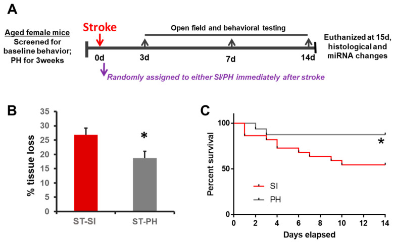
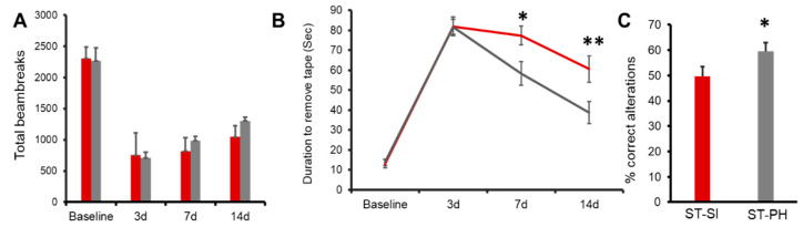
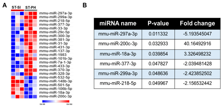
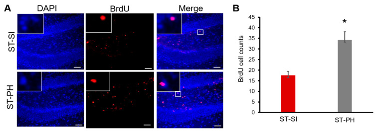
Social isolation and loneliness are risk factors for stroke. Elderly women are more likely to be isolated. Census data shows that in homeowners over the age of 65, women are much more likely to live alone. However, the underlying mechanisms of the detrimental effects of isolation have not been well studied in older females. In this study, we hypothesized that isolation impairs post-stroke recovery in aged female mice, leading to dysregulated microRNAs (miRNAs) in the brain, including those previously shown to be involved in response to social isolation (SI). Aged C57BL/6 female mice were subjected to a 60-min middle cerebral artery occlusion and were randomly assigned to either single housing (SI) or continued pair housing (PH) immediately after stroke for 15 days. SI immediately after stroke led to significantly more brain tissue loss after stroke and higher mortality. Furthermore, SI significantly delayed motor and sensory recovery and worsened cognitive function, compared to PH. A decrease in cell proliferation was seen in the dentate gyrus of SI mice assessed by bromodeoxyuridine (BrdU) labeling. miRNAome data analysis revealed changes in several miRNAs in the brain, such as miR-297a-3p and miR-200c-3p, which are known to regulate pathways involved in cell proliferation. In conclusion, our data suggest that SI can lead to a poor post-stroke recovery in aged females and dysregulation of miRNAs and reduced hippocampal cell proliferation.

Keywords: aging; ischemic stroke; miRNA; neurogenesis; social isolation.


Conflict of interest statement

The authors declare no conflict of interest.

Figures

Figure 1
Figure 1
Social isolation immediately after stroke increases infarct size and mortality in aged female mice. (A) The experimental timeline illustrates the study design. Animals were screened for baseline behavior and immediately assigned to either pair-housing or isolation. The animals were randomly assigned and housed either in continued pair-housing or separated and housed singly (SI). Behavioral testing was performed on days 3, 7 and 14. Mice were euthanized on day 15 for infarct analysis. (B) Post-stroke SI (ST-SI) mice had significantly more atrophy compared to post-stroke pair-housed mice (ST-PH) (n = 9/group; * p = 0.024). Student’s t-test was used after the normality of data was confirmed. (C) The socially isolated mice had significantly higher mortality compared to pair-housed mice (n = 22 in SI; 16 PH initially. * p = 0.043). P value was obtained from the log-rank test.
Figure 2
Figure 2
Post-stroke recovery is worse in isolated aged female mice compared to pair-housed mice. (A) Repeated open field analysis revealed that SI mice (red) had slower recovery compared to PH mice (grey). Although the SI mice showed no significant difference compared to PH mice, on day 14, the PH mice had a trend towards better locomotor activity compared to the SI mice (n = 12–13/group). (B) SI mice (red) also showed an increase in latency in the adhesive tape removal time compared to PH mice (grey) on day 14. (n = 12–13/group; * p = 0.020 at day 7, ** p = 0.005 at day 14 (C) The SI mice had a lower percentage of complete alteration in the Y-maze compared to the PH mice). ST-PH mice had significantly more% correct alterations compared to ST-SI mice (n = 12–13/group; * p = 0.05). Data are presented as mean ± SEM. Repeated two-way ANOVA was used to account for within-subject correlation for the repeated measures (A and B). Student’s t-test was used for group comparisons (C).
Figure 3
Figure 3
SI after stroke induces significant changes in brain miRNA profiles compared to PH. (A) 17 significantly upregulated and 6 downregulated miRNAs in the PH group compared to SI mice were identified and plotted as a heat map. (B) Further miRNAome analysis, based on the p value of p < 0.05 and a minimum fold change of 2 or higher, allowed us to identify the differentially regulated brain miRNAs between SI and PH stroke groups.
Figure 4
Figure 4
PH aged females had higher cell proliferation compared to SI animals after stroke. (A) Representative images of the hippocampus of brain sections from mice administered bromodeoxyuridine (BrdU) (day 3 to 7) after stroke. Immunohistochemistry analysis and co-labeling with anti-BrdU (in Red) and DAPI (in blue) showed that mice that were isolated immediately after a stroke had lower BrdU-positive cells in the dentate gyrus. Magnified images in insert show co-labeling of BrdU and DAPI. Scale bar denotes 75 µm (B) Quantification of these BrdU-positive cells showed a significant increase in cell proliferation in the dentate gyrus of ST-PH mice compared to the ST-SI mice (n = 4/group. * p = 0.008). Data are presented as mean ± SEM. Student’s t-test was used for group comparisons.

References

    1. Jiaquan Xu M.D., Sherry L., Murphy B.S., Kenneth D., Kochanek M.A., Arias E. Mortality in the United States, 2018. In: United States Department of Health and Human Services, editor. Services. National Center for Health Statistics Hyattsville; Hyattsville, MD, USA: 2020.
    1. Wang J., Bai L., Shi M., Yang L., An Z., Li B., Zhao W., Gu H., Zhan C., Tu J., et al. Trends in Age of First-Ever Stroke Following Increased Incidence and Life Expectancy in a Low-Income Chinese Population. Stroke. 2016;47:929–935. doi: 10.1161/STROKEAHA.115.012466. - DOI - PubMed
    1. Valtorta N.K., Kanaan M., Gilbody S., Ronzi S., Hanratty B. Loneliness and social isolation as risk factors for coronary heart disease and stroke: Systematic review and meta-analysis of longitudinal observational studies. Heart. 2016;102:1009–1016. doi: 10.1136/heartjnl-2015-308790. - DOI - PMC - PubMed
    1. Rico-Uribe L.A., Caballero F.F., Martín-María N., Cabello M., Ayuso-Mateos J.L., Miret M. Association of loneliness with all-cause mortality: A meta-analysis. PLoS ONE. 2018;13:e0190033. doi: 10.1371/journal.pone.0190033. - DOI - PMC - PubMed
    1. Pantell M.S., Rehkopf D., Jutte D., Syme S.L., Balmes J., Adler N. Social Isolation: A Predictor of Mortality Comparable to Traditional Clinical Risk Factors. Am. J. Public Health. 2013;103:2056–2062. doi: 10.2105/AJPH.2013.301261. - DOI - PMC - PubMed