Review
. 2020 Dec 24;22(1):100.
doi: 10.3390/ijms22010100.

Myelin Oligodendrocyte Glycoprotein Antibody-Associated Disease: Current Insights into the Disease Pathophysiology, Diagnosis and Management

Affiliations
Review

Myelin Oligodendrocyte Glycoprotein Antibody-Associated Disease: Current Insights into the Disease Pathophysiology, Diagnosis and Management

Wojciech Ambrosius et al. Int J Mol Sci. .

Abstract

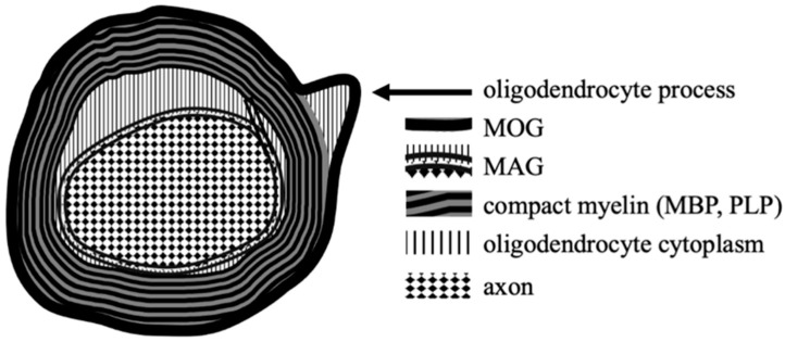
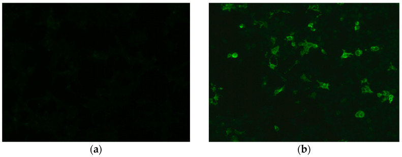
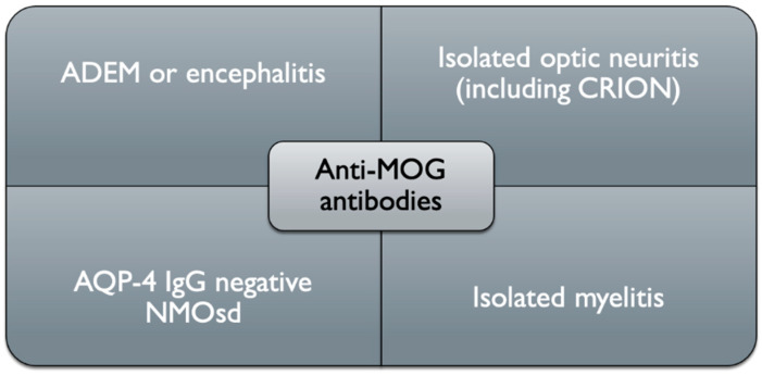
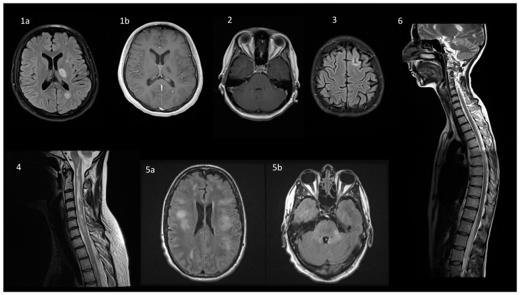
Myelin oligodendrocyte glycoprotein (MOG)-associated disease (MOGAD) is a rare, antibody-mediated inflammatory demyelinating disorder of the central nervous system (CNS) with various phenotypes starting from optic neuritis, via transverse myelitis to acute demyelinating encephalomyelitis (ADEM) and cortical encephalitis. Even though sometimes the clinical picture of this condition is similar to the presentation of neuromyelitis optica spectrum disorder (NMOSD), most experts consider MOGAD as a distinct entity with different immune system pathology. MOG is a molecule detected on the outer membrane of myelin sheaths and expressed primarily within the brain, spinal cord and also the optic nerves. Its function is not fully understood but this glycoprotein may act as a cell surface receptor or cell adhesion molecule. The specific outmost location of myelin makes it a potential target for autoimmune antibodies and cell-mediated responses in demyelinating processes. Optic neuritis seems to be the most frequent presenting phenotype in adults and ADEM in children. In adults, the disease course is multiphasic and subsequent relapses increase disability. In children ADEM usually presents as a one-time incident. Luckily, acute immunotherapy is very effective and severe disability (ambulatory and visual) is less frequent than in NMOSD. A critical element of reliable diagnosis is detection of pathogenic serum antibodies MOG with accurate, specific and sensitive methods, preferably with optimized cell-based assay (CBA). MRI imaging can also help in differentiating MOGAD from other neuro-inflammatory disorders. Reports on randomised control trials are limited, but observational open-label experience suggests a role for high-dose steroids and plasma exchange in the treatment of acute attacks, and for immunosuppressive therapies, such as steroids, oral immunosuppressants and rituximab as maintenance treatment. In this review, we present up-to-date clinical, immunological, radiographic, histopathological data concerning MOGAD and summarize the practical aspects of diagnosing and managing patients with this disease.

Keywords: NMO spectrum disorder; myelin oligodendrocyte glycoprotein (MOG); myelin oligodendrocyte glycoprotein associated disease (MOGAD); neuroimmunology; neuromyelitis optica (NMO).


Conflict of interest statement

All authors declare no conflict of interest.

Figures

Figure 1
Figure 1
The locations of MOG and other myelin proteins on oligodendrocytes within CNS.
Figure 2
Figure 2
Detection of MOG IgG in a cell-based assay: (a) negative control stain; (b) positive MOG IgG reaction. Images from the collection of Division of Neurochemistry and Neuropathology, Poznan University of Medical Sciences.
Figure 3
Figure 3
Schematic clinical representation of anti-MOG associated disorders (ADEM, acute disseminated encephalomyelitis, AQP-4, aquaporin-4, MOG, myelin oligodendrocyte glycoprotein, CRION, chronic relapsing inflammatory optic neuropathy, NMO, neuromyelitis optica).
Figure 4
Figure 4
Typical magnetic resonance findings in a spectrum of diseases overlapping with MOGAD. (1) ADEM presentation in a 17-year old female with behavioral changes and transient right-sided paresis. On axial FLAIR (1a), a periventricular lesion at the level of the left subcortical nuclei (17 × 11 mm) and the left ventricular triangle (15 × 10 mm) are visualized. On axial post-contrast T1-weighted image (1b) slight contrast enhancement is present in the latter. (2) Optic neuritis in a 26-year old female with AQP4-ab positive NMOsd: bilateral enhancement of the optic nerves on axial T1-weighted post-contrast sequence. (3) Cortical involvement in a 22-year old female with MS: on axial FLAIR there is a 4 mm long linear hyperintensity that surrounds the cortical gyrus and involves both, the cortex and subcortical white matter. (4) AQP4-ab positive NMOsd in a 43-year old female: on a sagittal T2-weighted sequence LETM is visualized from C1–C2 to C5. (5) MOGAD in a 47-year old male presenting with ADEM with confirmed MOG-ab seropositivity. On axial FLAIR images there are diffuse nonspecific white matter hyperintensities involving both cerebral hemispheres, corpus callosum (5a), posterior fossa (5b), but periventricular lesions are lacking. (6) MOGAD in a 34-year old female presenting with spastic paraparesis, which improved significantly after a 5-day intravenous methylprednisolone course: on a sagittal T2-weighted sequence multiple hyperintensive poorly demarcated lesions are visible throughout the spinal cord, including the cervical and thoracic regions, and affecting medullary conus, as well. The lesions are not visible on T1-weighted sequence and are non-enhancing. Abbreviations: MOGAD—MOG-antibody associated disease, ADEM—acute disseminated encephalomyelitis, FLAIR—Fluid Attenuated Inversion Recovery, NMOsd—neuromyelitis optica spectrum disorder, AQP4—aquaporin 4, LETM—longitudinally extensive transverse myelitis. Subfigures 1a,b, 2–4 and 6: from the collection of the Department of Neurology, Poznan University of Medical Sciences, Poznan, Poland. Subfigure 5a,b: courtesy of dr Claudia F. Lucchinetti, Department of Neurology, Mayo Clinic, Rochester, MN, USA

References

    1. Schumacher G.A., Beebe G., Kibler R.F., Kurland L.T., Kurtzke J.F., McDowell F., Nagler B., Sibley W.A., Tourtellotte W.W., Willmon T.L. Problems of experimental trials of therapy in multiple sclerosis: Report by the panel on the evaluation of experimental trials of therapy in multiple sclerosis. Ann. N. Y. Acad. Sci. 1965;122:552–568. doi: 10.1111/j.1749-6632.1965.tb20235.x. - DOI - PubMed
    1. Poser C.M., Paty D.W., Scheinberg L., McDonald W.I., Davis F.A., Ebers G.C., Johnson K.P., Sibley W.A., Silberberg D.H., Tourtellotte W.W. New diagnostic criteria for multiple sclerosis: Guidelines for research protocols. Ann. Neurol. 1983;1393:227–231. doi: 10.1002/ana.410130302. - DOI - PubMed
    1. Thompson A.J., Banwell B.L., Barkhof F., Carroll W.M., Coetzee T., Comi G., Correale J., Fazekas F., Filippi M., Freedman M.S., et al. Diagnosis of multiple sclerosis: 2017 revisions of the McDonald criteria. Lancet Neurol. 2018;17:162–173. doi: 10.1016/S1474-4422(17)30470-2. - DOI - PubMed
    1. Wingerchuk D.M., Banwell B., Bennett J.L., Cabre P., Carroll W., Chitnis T., De Seze J., Fujihara K., Greenberg B., Jacob A., et al. International consensus diagnostic criteria for neuromyelitis opstica spectrum disorders. Neurology. 2015;85:177–189. doi: 10.1212/WNL.0000000000001729. - DOI - PMC - PubMed
    1. Jasiak-Zatonska M., Kalinowska-Lyszczarz A., Michalak S., Kozubski W. The immunology of neuromyelitis optica—Current knowledge, clinical implications, controversies and future perspectives. Int. J. Mol. Sci. 2016;17:273. doi: 10.3390/ijms17030273. - DOI - PMC - PubMed

MeSH terms