. 2020 Dec 24;13(1):19.
doi: 10.3390/pharmaceutics13010019.

Electrochemotherapy with Calcium Chloride and 17β-Estradiol Modulated Viability and Apoptosis Pathway in Human Ovarian Cancer

Affiliations

Electrochemotherapy with Calcium Chloride and 17β-Estradiol Modulated Viability and Apoptosis Pathway in Human Ovarian Cancer

Zofia Łapińska et al. Pharmaceutics. .

Abstract

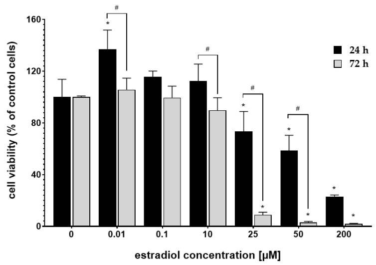
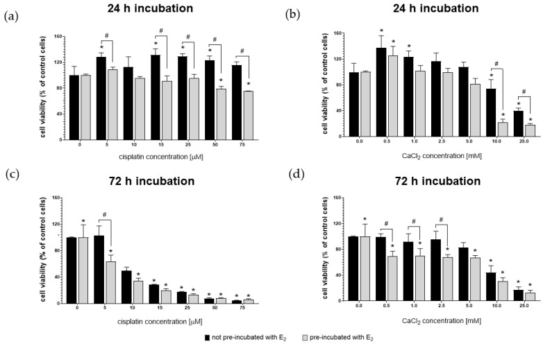
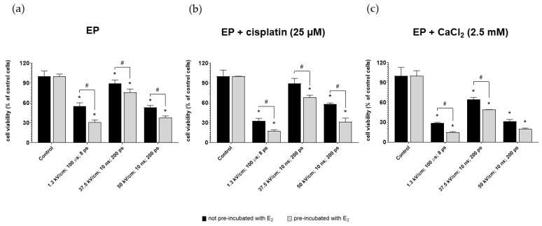
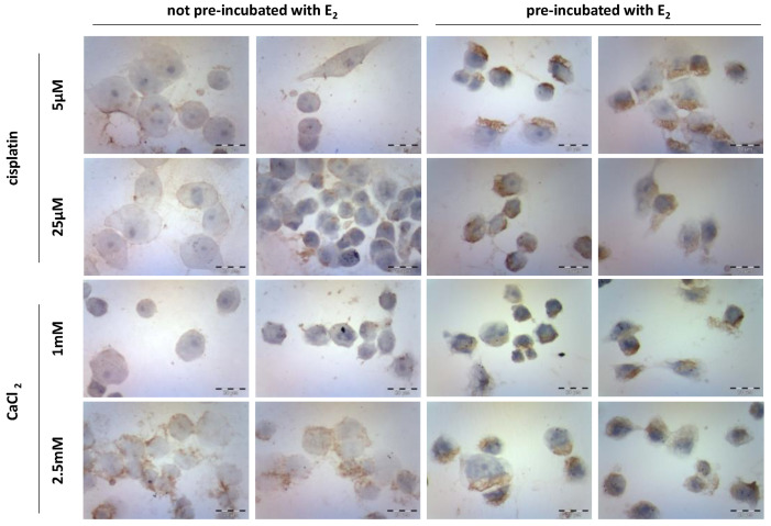
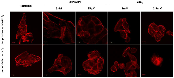
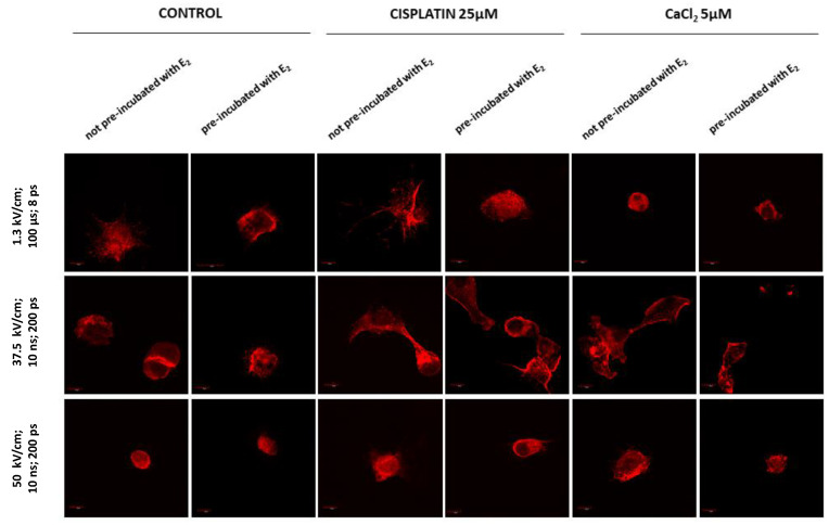
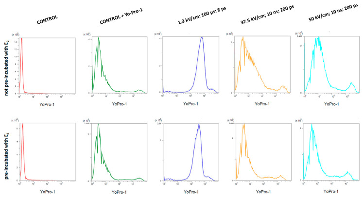
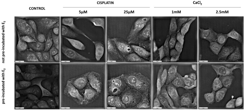
Estrogens (Es) play a significant role in the carcinogenesis and progression of ovarian malignancies. Depending on the concentration, Es may have a protective or toxic effect on cells. Moreover, they can directly or indirectly affect the activity of membrane ion channels. In the presented study, we investigated in vitro the effectiveness of the ovarian cancer cells (MDAH-2774) pre-incubation with 17β-estradiol (E2; 10 µM) in the conventional chemotherapy (CT) and electrochemotherapy (ECT) with cisplatin or calcium chloride. We used three different protocols of electroporation including microseconds (µsEP) and nanoseconds (nsEP) range. The cytotoxic effect of the applied treatment was examined by the MTT assay. We used fluorescent staining and holotomographic imaging to observe morphological changes. The immunocytochemical staining evaluated the expression of the caspase-12. The electroporation process's effectiveness was analyzed by a flow cytometer using the Yo-Pro™-1 dye absorption assay. We found that pre-incubation of ovarian cancer cells with 17β-estradiol may effectively enhance the chemo- and electrochemotherapy with cisplatin and calcium chloride. At the same time, estradiol reduced the effectiveness of electroporation, which may indicate that the mechanism of increasing the effectiveness of ECT by E2 is not related to the change of cell membrane permeability.

Keywords: 17β-estradiol; MDAH-2774; calcium electroporation; chemotherapy; electrochemotherapy; ovarian cancer.


Conflict of interest statement

The authors declare that they have no conflict of interest concerning this article.

Figures

Figure 1
Figure 1
MDAH-2774 cells viability measured by the MTT assay after 24 h and 72 h incubation in different 17β-estradiol concentrations. Notes: (mean ± SD) N = 3, * p < 0.05 compared to control, # p < 0.05 compared between different incubation times.
Figure 2
Figure 2
MDAH-2774 cells viability measured by the MTT assay after 24 h (a,b) and 72 h (c,d) incubation in increasing cisplatin (a,c) and CaCl2 (b,d) concentrations. Notes: (mean ± SD) N = 3, * p < 0.05 compared to control, # p < 0.05 compared between different pre-incubation conditions.
Figure 3
Figure 3
MDAH-2774 cells viability measured by the MTT assay after (a) µEP and nsEP with (b) 25 µM cisplatin and (c) 2.5 mM CaCl2. Notes: (mean ± SD) N = 3, * p < 0.05 compared to control, # p < 0.05 compared between different pre-incubation conditions; ps—pulses.
Figure 4
Figure 4
Caspase-12 immunocytochemical staining after exposure to cisplatin (5 µM and 25 µM) and CaCl2 (1 mM and 2.5 mM) in the MDAH-2774 cell line.
Figure 5
Figure 5
Immunofluorescence staining of MDAH-2774 cells (60×) after 24 h incubation with cisplatin and CaCl2. Alexa FluorTM 546 phalloidin used for actin filaments labeled. Scale bars correspond to 100 µm.
Figure 6
Figure 6
Immunofluorescence staining of MDAH-2774 cells (60×) after µEP and nsEP with the addition of cisplatin and CaCl2. Alexa FluorTM 546 phalloidin used for actin filaments labeled. Scale bars represent 100 µm; ps—pulses.
Figure 7
Figure 7
Yo-Pro-1TM fluorescent dye absorption in MDAH-2774 cells after µEP and nsEP; ps—pulses.
Figure 8
Figure 8
Holotomographic microscopy studies after 24 h with cisplatin and CaCl2. Scale bars represent 20 µM.

References

    1. Coburn S.B., Bray F., Sherman M.E., Trabert B. International patterns and trends in ovarian cancer incidence, overall and by histologic subtype. Int. J. Cancer. 2017;140:2451–2460. doi: 10.1002/ijc.30676. - DOI - PMC - PubMed
    1. Bray F., Ferlay J., Soerjomataram I., Siegel R.L., Torre L.A., Jemal A. Global cancer statistics 2018: GLOBOCAN estimates of incidence and mortality worldwide for 36 cancers in 185 countries. CA Cancer J. Clin. 2018;68:394–424. doi: 10.3322/caac.21492. - DOI - PubMed
    1. Huang Z., Yan H., Chavan D., Yuan Z., Yang X., Zhang Y., Song K., Kong B. Effective treatment of a patient with stage IV ovarian cancer: A case report. Oncol. Lett. 2018;15:588–591. - PMC - PubMed
    1. Stewart C., Ralyea C., Lockwood S. Ovarian Cancer: An Integrated Review. Semin. Oncol. Nurs. 2019;35:151–156. doi: 10.1016/j.soncn.2019.02.001. - DOI - PubMed
    1. Mungenast F., Thalhammer T. Estrogen Biosynthesis and Action in Ovarian Cancer. Front. Endocrinol. 2014;5:1–12. doi: 10.3389/fendo.2014.00192. - DOI - PMC - PubMed

LinkOut - more resources