. 2020 Dec 24;11(1):22.
doi: 10.3390/nano11010022.

Bone Regeneration Capacity of Newly Developed Uncalcined/Unsintered Hydroxyapatite and Poly-l-lactide-co-glycolide Sheet in Maxillofacial Surgery: An In Vivo Study

Affiliations

Bone Regeneration Capacity of Newly Developed Uncalcined/Unsintered Hydroxyapatite and Poly-l-lactide-co-glycolide Sheet in Maxillofacial Surgery: An In Vivo Study

Huy Xuan Ngo et al. Nanomaterials (Basel). .

Abstract

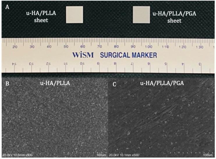
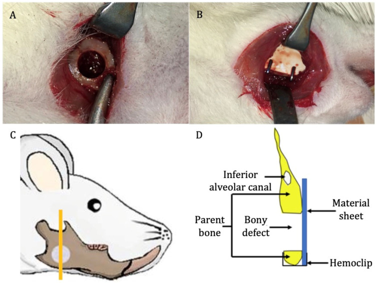
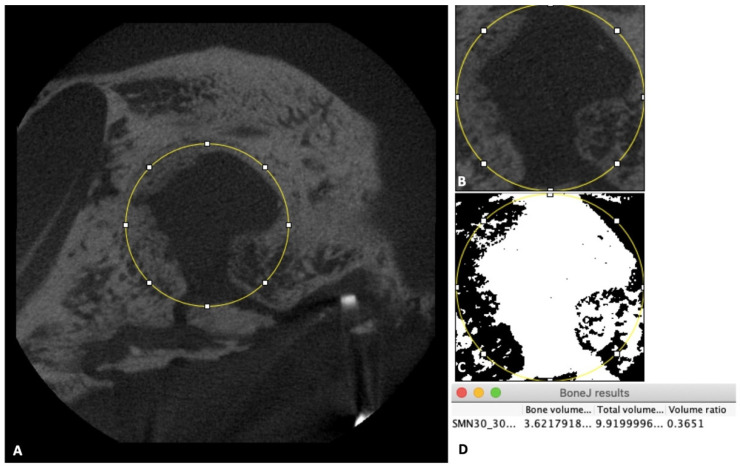
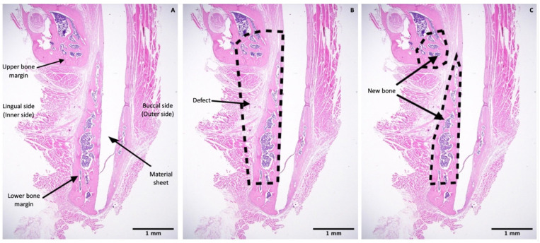
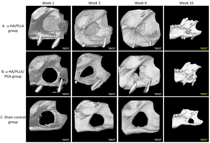
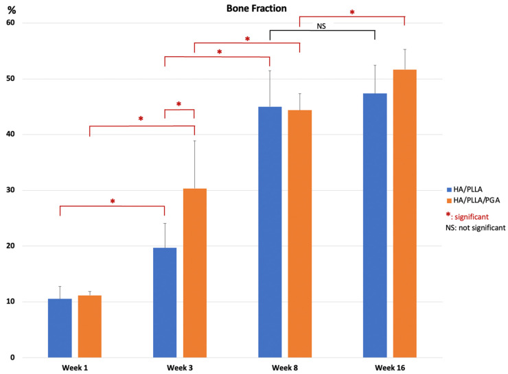
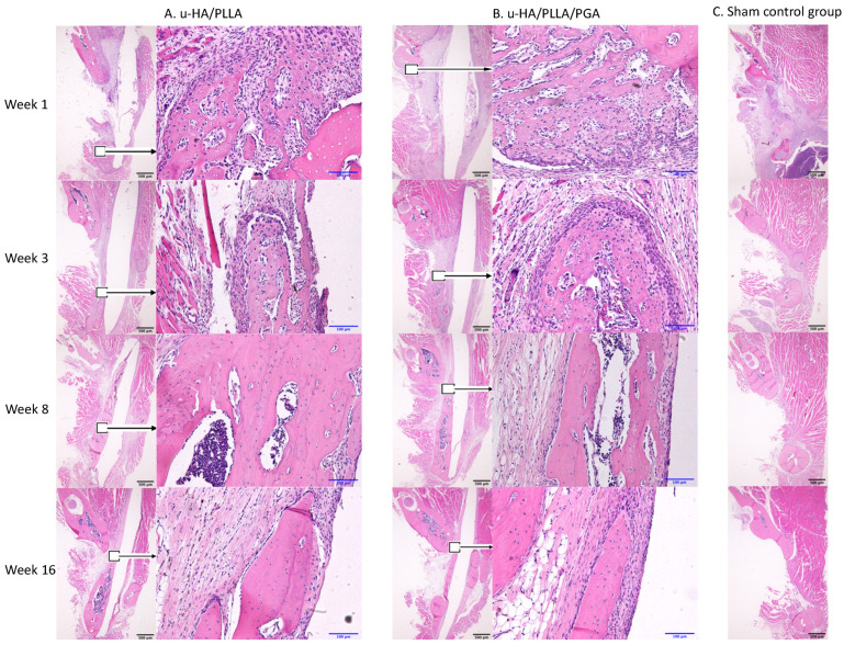
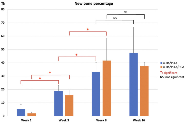
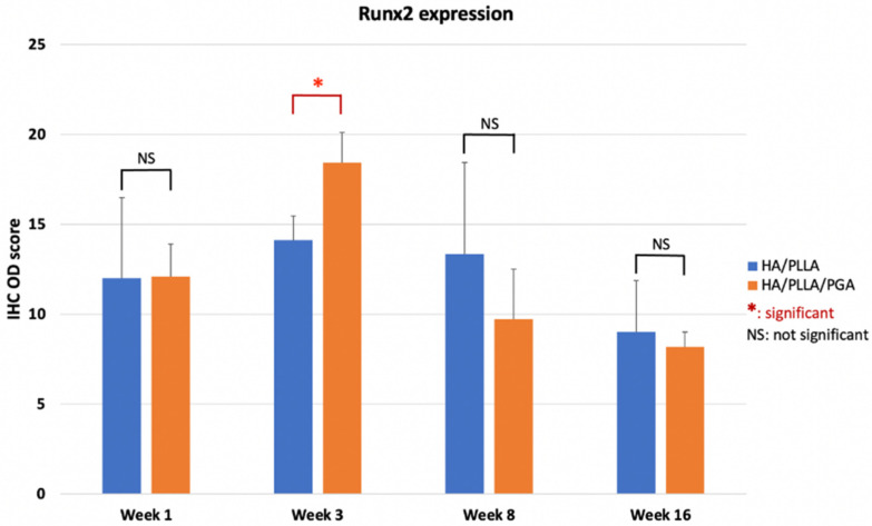
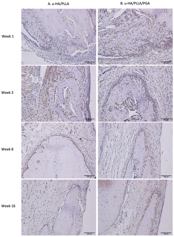
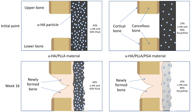
Uncalcined/unsintered hydroxyapatite and poly-l-lactide-co-glycolide (u-HA/PLLA/PGA) is a new bioresorbable nanomaterial with superior characteristics compared with current bioresorbable materials, including appropriate mechanical properties, outstanding bioactive/osteoconductive features, and remarkably shorter resorption time. Nevertheless, the bone regeneration characteristics of this nanomaterial have not been evaluated in maxillofacial reconstructive surgery. In this study, we used a rat mandible model to assess the bone regeneration ability of u-HA/PLLA/PGA material, compared with uncalcined/unsintered hydroxyapatite and poly-l-lactide acid (u-HA/PLLA) material, which has demonstrated excellent bone regenerative ability. A 4-mm-diameter defect was created at the mandibular angle area in 28 Sprague Dawley male rats. The rats were divided into three groups: u-HA/PLLA/PGA (u-HA/PLLA/PGA graft + defect), u-HA/PLLA (u-HA/PLLA graft + defect), and sham control (defect alone). At 1, 3, 8, and 16 weeks after surgeries, the rats were sacrificed and assessed by micro-computed tomography, histological analysis with hematoxylin and eosin staining, and immunohistochemical analyses. The results confirmed that the accelerated bone bioactive/regenerative osteoconduction of u-HA/PLLA/PGA was comparable with that of u-HA/PLLA in the rat mandible model. Furthermore, this new regenerative nanomaterial was able to more rapidly induce bone formation in the early stage and had great potential for further clinical applications in maxillofacial reconstructive surgery.

Keywords: Runx2; bone regeneration; leptin receptor; osteocalcin; osteoconductivity; poly-l-lactic acid; poly-l-lactide-co-glycolide; uncalcined/unsintered hydroxyapatite.


Conflict of interest statement

The authors have no conflict of interest to declare.

Figures

Figure 1
Figure 1
Materials. (A) Examples of a u-HA/PLLA (left) and u-HA/PLLA/PGA (right) sheets. (B) Scanning electron microscope image of u-HA/PLLA material. (C) Scanning electron microscope image of u-HA/PLLA/PGA material.
Figure 2
Figure 2
Surgical procedure. (A) Critical-size defect created at the mandibular angle on the right side. (B) Placement of reconstructive material. (C) Site of sample taken for analysis (the orange vertical line) (image modified from Sha et al. [18]). (D) Schematic coronal view of the specimen.
Figure 3
Figure 3
Micro-CT evaluation. (A) Image of the 4-mm-diameter circle including the initial defect and new bone inside the defect. (B) Illustration of the new 3D volume after using the “Duplicate” tool. (C) Illustration of the new 3D volume after using the “Make binary” tool. (D) Box shows final results.
Figure 4
Figure 4
Histomorphometric evaluation. (A) Image at 1.25× magnification including the upper and lower bony margins of the defect region and the sheet covering the defect. (B) Illustration of the whole defect region. (C) Illustration of the new bone area in upper and lower bony margins. Scale bars: 1 mm (Black).
Figure 5
Figure 5
IHC OD score for LepR staining. (A) Illustration of a positive image at 100× magnification. (B) Positive score for image A after using the IHC profiler plugin in Fiji software. (C) Illustration of negative image at 100× magnification. (D) Negative score for image C after using the IHC profiler plugin in Fiji software.
Figure 6
Figure 6
Example of new bone area ROI selection (anti-OCN IHC staining). (A) Original image with overlay of the selected ROI, which is the area of new bone. *, selected area; +, unselected area. (B) Hematoxylin-stained image separated from the original image. (C) DAB-stained image separated from the original image. (D) Superimposition of the saved ROI onto the DAB-stained image. *, selected area; +, unselected area.
Figure 7
Figure 7
3D micro-CT images. (A) 3D views of the u-HA/PLLA group. (B) 3D views of the u-HA/PLLA/PGA group. Newly formed bone increased over time. (C) 3D views of the sham control group. New bone formation could not be identified. To present a broad view, images at week 16 are lower magnification than images at other weeks. Scale bars: 2000 μm (white), 5000 μm (yellow).
Figure 8
Figure 8
Percentage of new bone volume determined by micro-CT evaluation of u-HA/PLLA and u-HA/PLLA/PGA groups. * p < 0.05.
Figure 9
Figure 9
Hematoxylin and eosin-stained sections from the u-HA/PLLA, u-HA/PLLA/PGA, and sham control groups at weeks 1, 3, 8, and 16. Images in each subgroup were taken at 1.25× and 20× magnification (from left to right). Images of the sham control group were taken at 1.25× magnification. (A) u-HA/PLLA group. (B) u-HA/PLLA/PGA. (C) Sham control group. Scale bars: 100 μm (blue), 500 μm (black).
Figure 10
Figure 10
Percentages of new bone area in histomorphometric evaluation of u-HA/PLLA and u-HA/PLLA/PGA groups. * p < 0.05.
Figure 11
Figure 11
IHC OD score of Runx2 in the u-HA/PLLA and u-HA/PLLA/PGA groups. * p < 0.05.
Figure 12
Figure 12
Runx2 expression in the u-HA/PLLA and u-HA/PLLA/PGA groups. All images were taken at 20× magnification. Runx2 expression was similar in the u-HA/PLLA and u-HA/PLLA/PGA groups. (A) u-HA/PLLA group. (B) u-HA/PLLA/PGA group. Scale bar: 100 μm (black).
Figure 13
Figure 13
LepR expression in the u-HA/PLLA and u-HA/PLLA/PGA groups. All images were taken at 20× magnification. (A) u-HA/PLLA group. (B) u-HA/PLLA/PGA group. Scale bar: 100 μm (black).
Figure 14
Figure 14
IHC OD score of LepR in the u-HA/PLLA and u-HA/PLLA/PGA groups. * p < 0.05.
Figure 15
Figure 15
OCN expression in the u-HA/PLLA and u-HA/PLLA/PGA groups. All images were taken at 1.25× magnification. (A) u-HA/PLLA group. (B) u-HA/PLLA/PGA group. Scale bar: 500 μm (black).
Figure 16
Figure 16
Digital H-scores based on IHC staining with anti-OCN antibody in the u-HA/PLLA and u-HA/PLLA/PGA groups. * p < 0.05.
Figure 17
Figure 17
Theoretical explanation of the relationships between the bone remodeling capacity of u-HA/PLLA/PGA material and the u-HA particle proportion and degradation rate. Because of the more rapid degradation process, the u-HA particles in the new nanomaterial may be exposed to body fluids at an earlier stage.

Similar articles

Cited by

References

    1. Tschakaloff A., Losken H.W., Lalikos J., Link J., Mooney M.P., von Oepen R., Michaeli W., Losken A. Experimental studies of DL-polylactic acid biodegradable plates and screws in rabbits: Computed tomography and molecular weight loss. J. Craniofac. Surg. 1993;4:223–227. - PubMed
    1. Yamamuro T., Matsusue Y., Uchida A., Shimada K., Shimozaki E., Kitaoka K. Bioabsorbable osteosynthetic implants of ultra high strength poly-L-lactide. A clinical study. Int. Orthop. 1994;18:332–340. doi: 10.1007/BF00187076. - DOI - PubMed
    1. Rokkanen P.U. Absorbable materials in orthopaedic surgery. Ann. Med. 1991;23:109–115. doi: 10.3109/07853899109148033. - DOI - PubMed
    1. Goldstein J.A. The use of bioresorbable material in craniofacial surgery. Clin. Plast. Surg. 2001;28:653–659. doi: 10.1016/S0094-1298(20)32413-5. - DOI - PubMed
    1. Sanger C., Soto A., Mussa F., Sanzo M., Sardo L., Donati P.A., Di Pietro G., Spacca B., Giordano F., Genitori L. Maximizing results in craniofacial surgery with bioresorbable fixation devices. J. Craniofac. Surg. 2007;18:926–930. doi: 10.1097/scs.0b013e3180a771e9. - DOI - PubMed

LinkOut - more resources