Review
. 2020 Dec 24;10(1):11.
doi: 10.3390/antiox10010011.

Paraoxonase Role in Human Neurodegenerative Diseases

Affiliations
Review

Paraoxonase Role in Human Neurodegenerative Diseases

Cadiele Oliana Reichert et al. Antioxidants (Basel). .

Abstract

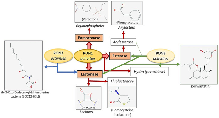
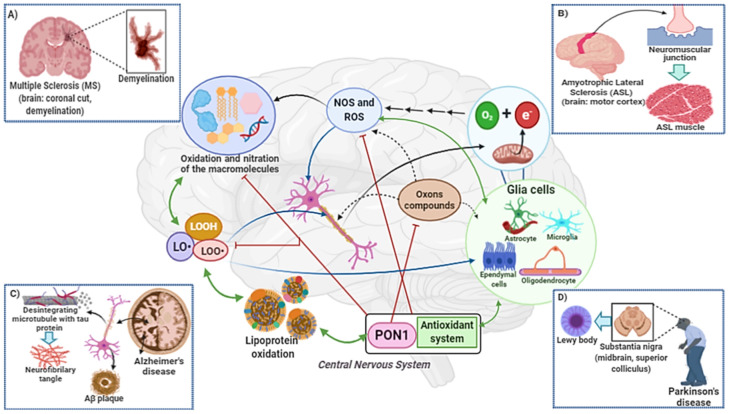
The human body has biological redox systems capable of preventing or mitigating the damage caused by increased oxidative stress throughout life. One of them are the paraoxonase (PON) enzymes. The PONs genetic cluster is made up of three members (PON1, PON2, PON3) that share a structural homology, located adjacent to chromosome seven. The most studied enzyme is PON1, which is associated with high density lipoprotein (HDL), having paraoxonase, arylesterase and lactonase activities. Due to these characteristics, the enzyme PON1 has been associated with the development of neurodegenerative diseases. Here we update the knowledge about the association of PON enzymes and their polymorphisms and the development of multiple sclerosis (MS), amyotrophic lateral sclerosis (ALS), Alzheimer's disease (AD) and Parkinson's disease (PD).

Keywords: Alzheimer’s disease; Parkinson’s disease; amyotrophic lateral sclerosis; multiple sclerosis; oxidative stress; paraoxonases.


Conflict of interest statement

The authors declare no conflict of interest.

Figures

Figure 1
Figure 1
First, dietary fat is emulsified by bile salts, being degraded by lipases. Fatty acids are absorbed through the intestinal mucosa, being converted into triacylglycerol. Then there is the joint incorporation of cholesterol, triglycerides and apolipoproteins (Apo-CII and Apo-B48), forming the chylomicrons. Dietary fat is transported in the blood and lymph vessels by the chylomicrons. Apo-CII on the surface of chylomicron activates the enzyme lipoprotein lipase (LPL), which releases fatty acids and glycerol to tissues, mainly muscle and adipose tissues. In tissues, fatty acids can be esterified and stored or used to generate energy in cellular function. This process results in the formation of chylomicron remnants (Apo-B48 and Apo-E in surface), that have cholesterol and low triglyceride content. They are transported to the liver, where receptors bind to the Apo-E, leading to endocytosis and subsequent degradation in lysosomes, with the release of cholesterol and fatty acids. In the liver, very-low density lipoprotein (VLDL) is formed. It is rich in triacylglycerols (about 55%) and contains on its surface the following Apos: B100, E and C I, II, III. The Apo-C and -E in VLDL come from plasma high-density lipoproteins (HDL). In addition, during the lipolysis process, Apo-C, cholesterol and phospholipids are transferred from VLDL to HDL. LPL hydrolyzes VLDL in capillaries releasing fatty acids to tissues. The hydrolysis process gives rise to the IDL (or remaining VLDL), with Apo-E and Apo-B100 on its surface. IDLs are endocytosed in the liver, after binding to the low-density lipoprotein (LDL) (Apo-B100) or LDL receptor related protein (LRP) (binding to the Apo-E). IDL is also converted to LDL (about 50% cholesterol) by the action of liver LPL, which contains only one Apo-B100 molecule on its surface. This mechanism is not yet fully described. However, it is believed that there is an exchange of apolipoproteins and lipid content between HDL and IDL, by cholesterol ester transfer protein (CETP), together with phospholipid and triacylglycerol hydrolysis by hepatic LPL for the formation of LDL. LDL transports cholesterol to the extra-hepatic tissues by the binding of Apo-B100 to the LDL receptor or scavenger receptors followed by endocytosis. LDL receptor expression is downregulated by the content of intracellular cholesterol. Cholesterol is transported from tissues to the liver by HDL, which contains apolipoproteins A, C and E. In tissues, the ATP-binding cassette (ABC)-A1 and G1 proteins participate of the transport of cholesterol to HDL. Cholesterol in plasma is esterified by lecithin-cholesterol acyl transferase (LCAT), an enzyme activated by apolipoprotein-AI (Apo-AI). LCAT, in conjunction with lecithin (present in HDL), catalyzes the formation of cholesterol esters from fatty acids. As HDL captures cholesterol from tissues or lipoprotein hydrolysis, its diameter increases, from nascent HDL to discoid HDL. When the cholesterol ester content is taken, HDL migrates to the liver, binds to the Apo-AI receptor, and is released in hepatocytes. Then, cholesterol can originate VLDL or be excreted via the biliary or fecal pathways.
Figure 2
Figure 2
Paraoxonase family activities and substrates.
Figure 3
Figure 3
HDL biosynthesis occurs in the liver and in a small part of the intestine. Paraoxonase 1 (PON1) biosynthesis occurs only in the liver. After the formation of both HDL and PON1, they enter into the circulation. An association between HDL and PON1 can occur both in the hepatic circulation (to a lesser extent) and in the plasma. A small amount is free PON1, which do not adhere to any lipoprotein. An important function of PON1 is to prevent oxidation of both HDL and LDL, through the hydrolysis of reactive compounds. Oxidized LDL (ox-LDL) is pro-inflammatory and atherogenic. PON1 is carried to the tissues by HDL, where it performs its function as an antioxidant enzyme. In addition, the portion of free PON1 in the plasma also acts as an antioxidant, but its hydrolysis capacity is reduced.
Figure 4
Figure 4
The consumption of oxygen by the brain is high. O2 is used in oxidative phosphorylation to produce energy in the form of ATP in mitochondria. ATP is used to maintain membrane potential, the flow of neurotransmitters and the synapse in neurons. During the ATP synthesis process, occurs electron (e-) leakage of the electron transport chain. The free electron reacts with oxygen and forms the superoxide radical (O2•−). Then, hydroxyl radical formation can occur from the Fenton and Haber–Weiss reactions. Reactive oxygen species (ROS) and reactive nitrogen species (NOS) react with macromolecules: Lipids, proteins, carbohydrates and DNA. Lipid peroxidation of polyunsaturated fatty acids results in the accumulation of lipid hydroperoxides (LOOH). In the degradation of LOOH, the formation of alkoxyl (LO•) and peroxyl (LOO•) radicals occurs, highly reactive with proteins and lipid members. This process is widespread and can occur anywhere in the brain. Initially, it changes neuronal and glial cell function, and then cell death. In addition, in the lipid peroxidation process cytotoxic aldehydes are formed. Cellular metabolism generates O2•−, H2O2 and NO•, that can react and oxidize neurotransmitters. Under physiological conditions, the CNS antioxidant systems (superoxide dismutase (SOD), catalase (CAT), glutathione (GSH) and PON1) are able to maintain tissue homeostasis. However, when dyshomeostasis occurs, either by an increase in toxic substances (such as organophosphate compounds) and/or oxidative stress, dysregulating lipid homeostasis, then the neurodegeneration process begins. Increased oxidative stress and decreased activity of antioxidant enzymes, such as PON1, are strongly associated with the pathophysiology of multiple sclerosis (A), amyotrophic lateral sclerosis (B), Alzheimer’s disease (C) and Parkinson’s disease (D).

References

    1. World Health Organization . Risk Reduction of Cognitive Decline and Dementia: WHO Guidelines. WHO; Geneva, Switzerland: 2019. - PubMed
    1. WHO . Global Action Plan on the Public Health Response to Dementia 2017–2025. WHO; Geneva, Switzerland: 2017.
    1. Mathieu C., Pappu R.V., Paul Taylor J. Beyond aggregation: Pathological phase transitions in neurodegenerative disease. Science. 2020;370:56–60. doi: 10.1126/science.abb8032. - DOI - PMC - PubMed
    1. Reichert C.O., de Freitas F.A., Sampaio-Silva J., Rokita-Rosa L., de Barros P.L., Levy D., Bydlowski S.P. Ferroptosis mechanisms involved in neurodegenerative diseases. Int. J. Mol. Sci. 2020;21:8765. doi: 10.3390/ijms21228765. - DOI - PMC - PubMed
    1. Singh A., Kukreti R., Saso L., Kukreti S. Oxidative stress: A key modulator in neurodegenerative diseases. Molecules. 2019;24:1583. - PMC - PubMed

LinkOut - more resources