. 2020 Dec 24;26(1):53.
doi: 10.3390/molecules26010053.

Betulinic Acid Protects DOX-Triggered Cardiomyocyte Hypertrophy Response through the GATA-4/Calcineurin/NFAT Pathway

Affiliations

Betulinic Acid Protects DOX-Triggered Cardiomyocyte Hypertrophy Response through the GATA-4/Calcineurin/NFAT Pathway

Jung Joo Yoon et al. Molecules. .

Abstract

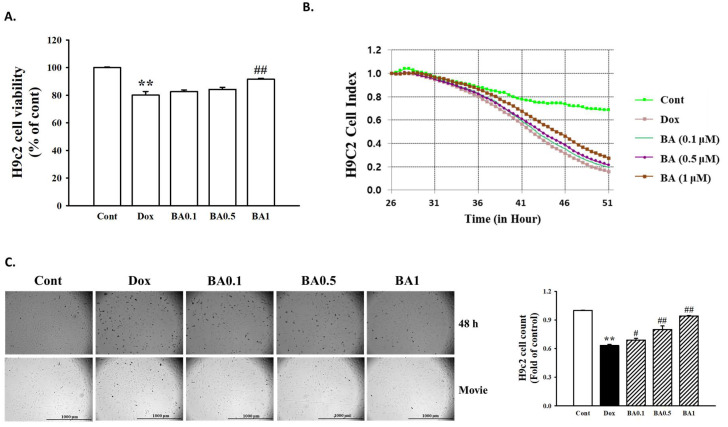
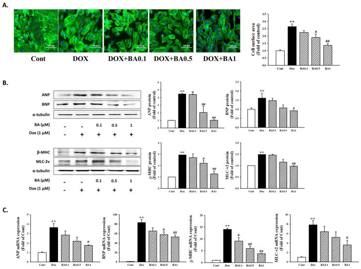
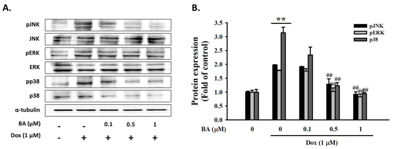
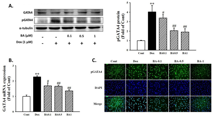
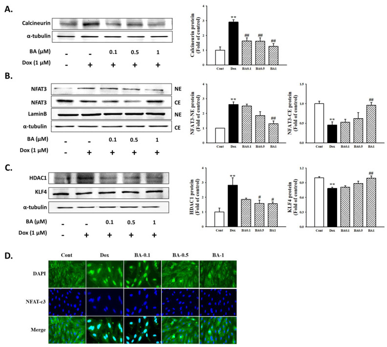
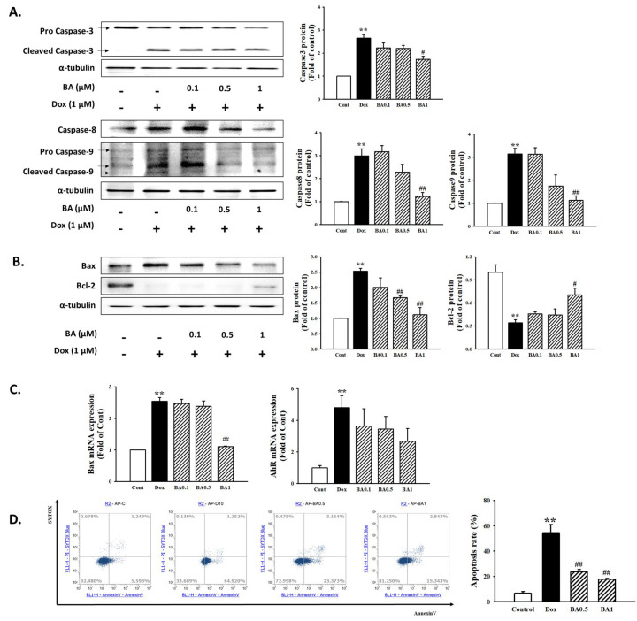
Cardiac hypertrophy is a major risk factor for heart failure and leads to cardiovascular morbidity and mortality. Doxorubicin (DOX) is regarded as one of the most potent anthracycline antibiotic agents; however, its clinical usage has some limitations because it has serious cardiotoxic side effects such as dilated cardiomyopathy and congestive heart failure. Betulinic acid (BA) is a pentacyclic-cyclic lupane-type triterpene that has been reported to have anti-bacterial, anti-inflammatory, anti-vascular neogenesis, and anti-fibrotic effects. However, there is no study about its direct effect on DOX induced cardiac hypertrophy and apoptosis. The present study aims to investigate the effect of BA on DOX-induced cardiomyocyte hypertrophy and apoptosis in vitro in H9c2 cells. The H9c2 cells were stimulated with DOX (1 µM) in the presence or absence of BA (0.1-1 μM) and incubated for 24 h. The results of the present study indicated that DOX induces the increase cell surface area and the upregulation of hypertrophy markers including atrial natriuretic peptide (ANP), B-type natriuretic peptide (BNP), beta-myosin heavy chain (β-MHC), and Myosin Light Chain-2 (MLC2) in H9c2 cells. However, the pathological hypertrophic responses were downregulated after BA treatment. Moreover, phosphorylation of JNK, ERK, and p38 in DOX treated H9c2 cells was blocked by BA. As a result of measuring the change in ROS generation using DCF-DA, BA significantly inhibited DOX-induced the production of intracellular reactive oxygen species (ROS) when BA was treated at a concentration of over 0.1 µM. DOX-induced activation of GATA-4 and calcineurin/NFAT-3 signaling pathway were remarkably improved by pre-treating of BA to H9c2 cells. In addition, BA treatment significantly reduced DOX-induced cell apoptosis and protein expression levels of Bax and cleaved caspase-3/-9, while the expression of Bcl-2 was increased by BA. Therefore, BA can be a potential treatment for cardiomyocyte hypertrophy and apoptosis that lead to sudden heart failure.

Keywords: apoptosis; betulinic acid; cardiomyocyte hypertrophy; doxorubicin; heart failure.


Conflict of interest statement

All authors declare that they have no potential conflicts of interest concerning this article.

Figures

Figure 1
Figure 1
Effect of betulinic acid (BA) on doxorubicin (DOX)-induced H9c2 cell death. (A) Effects of BA on DOX-induced changes of cell viability. H9c2 cells were pretreatment with different concentrations of BA for 30 min prior to DOX stimulation, and the cell viability was detected by 3-(4,5-dimethylthiazol-2-yl)-2,5-diphenyl tetrazolium (MTT) assay. (B) The results of H9c2 cell Index using xCELLigence RTCA DP Real time cell analyzer. (C) Observation of H9c2 live cell imaging under the Lionheart FX Automated Microscope (Scale bar = 1000 μm). Representative images and quantitative results demonstrating that BA (0.1–1 μM) inhibited DOX (1 μM; 48 h)-induced H9c2 cells. All experiments were performed at least three times. Data represent mean ± SD. ** p < 0.01, vs. control; # p < 0.05, ## p < 0.01, vs. DOX-treated cells.
Figure 2
Figure 2
The effects of BA on DOX-induced cardiac hypertrophy. (A) The effects of BA on cell surface area size. The cell surface area was measured using anti-F-actin staining (green) under fluorescence microscopy. The nucleus was stained with DAPI (blue) (Scale bar = 100 μm). (B,C) Effect of BA on cardiomyocyte hypertrophy markers in DOX-treated H9c2 cells. The relative expression of ANP, BNP, β-MHC, and MLC-2v was determined using Western blot analysis and RT-qPCR assay. All experiments were performed at least three times. Data represent mean ± SD. ** p < 0.01, vs. control; # p < 0.05, ## p < 0.01 vs. DOX-treated cells.
Figure 3
Figure 3
The effects of BA on the expression of MAPK markers. Protein expression of MAPK markers (JNK, ERK, and p38) analyzed by Western blot. (A) Representative blots and (B) quantitative results demonstrating that BA decreased the phosphorylated levels of JNK, ERK, and p38 in response to DOX. The results are expressed as the mean ± SE values of three experiments. ** p < 0.01, vs. control; ## p < 0.01, vs. DOX-treated cells.
Figure 4
Figure 4
Effect of BA on ROS formation in DOX-induced cardiac hypertrophy. (A) Analysis of ROS formation using DCFH-DA staining on H9c2 cells treated DOX and pre-administration of BA after 30 min of treatment. (B) Alternatively, the relative ROS levels were quantified by fluorescence microplate. The images were taken using light (magnification 20×). All experiments were performed at least three times. Data represent mean ± SD. ** p < 0.01, vs. control; # p < 0.05, ## p < 0.01, vs. DOX-treated cells.
Figure 5
Figure 5
The effects of BA on activation of GATA4. (A) The protein levels of GATA-4 and phosphorylated GATA-4 were determined by Western blot analysis. (B) GATA-4 mRNA level was analyzed by using Real-time PCR. (C) Immunofluorescent images of p-GATA-4 nuclear translocation under the laser scanning confocal microscopy were show (magnification 400×). Nuclei were stained with DAPI (blue) and p-GATA-4 was stained with Alexa Fluor 488 (green) (Immunofluorescence, 200×). The results are expressed as the mean ± SE values of three experiments. ** p < 0.01, vs. control; # p < 0.05, ## p < 0.01, vs. DOX-treated cells.
Figure 6
Figure 6
The effects of BA on calcineurin/NFAT-3 signaling pathway. Expression of calcineurin (A), nuclear localization of NFAT-3 (B), and HDAC1/KLF4 (C), were determined by Western blot analysis. (D) Nuclear translocation of NFAT was examined by immunofluorescence analysis (Immunofluorescence, 400×). The results are expressed as the mean ± SE values of three experiments. ** p < 0.01, vs. control; # p < 0.05, ## p < 0.01, vs. DOX-treated cells.
Figure 7
Figure 7
Effect of BA on cell apoptosis in DOX-induced cardiac hypertrophy. (A) Western blot analysis results showing the expression levels of apoptosis-related proteins including Caspase-3 and Caspase-9 in DOX-induced H9c2 cells. (B) The protein and mRNA expression of Bax and Bcl-2 was examined by Western blot analysis. (C) Bax and AhR mRNA expression was examined by real time PCR (D) Cell apoptosis was measured using Annexin-V/PI staining and flow cytometry. All experiments were performed at least three times. Data represent mean ± SD. ** p < 0.01, vs. control; # p < 0.05, ## p < 0.01, vs. DOX-treated cells.

References

    1. Cortes-Funes H., Coronado C. Role of anthracyclines in the era of targeted therapy. Cardiovasc. Toxicol. 2007;7:56–60. doi: 10.1007/s12012-007-0015-3. - DOI - PubMed
    1. Yanti O., Carlo G.T., Kathleen L.G., Janssens S., Crijns H.J., Moens A.L. Doxorubicin-induced cardiomyopathy: From molecular mechanisms to therapeutic strategies. J. Mol. Cell. Cardiol. 2012;52:1213–1225. - PubMed
    1. Armenian S.H., Lacchetti C., Lenihan D. Prevention and monitoring of cardiac dysfunction in survivors of adult cancers: American Society of Clinical Oncology Clinical Practice Guideline. J. Oncol. Pract. 2017;35:893–911. - PubMed
    1. Vejpongsa P., Yeh E.T. Prevention of anthracycline-induced cardiotoxicity: Challenges and opportunities. J. Am. Coll. Cardiol. 2014;64:938–945. doi: 10.1016/j.jacc.2014.06.1167. - DOI - PubMed
    1. Molkentin J.D. Calcineurin–NFAT signaling regulates the cardiac hypertrophic response in coordination with the MAPKs. Cardiovasc. Res. 2004;63:467–475. doi: 10.1016/j.cardiores.2004.01.021. - DOI - PubMed

MeSH terms