

Why Venous Leg Ulcers Have Difficulty Healing: Overview on Pathophysiology, Clinical Consequences, and Treatment
- PMID: 33374372
- PMCID: PMC7795034
- DOI: 10.3390/jcm10010029
Why Venous Leg Ulcers Have Difficulty Healing: Overview on Pathophysiology, Clinical Consequences, and Treatment
Abstract
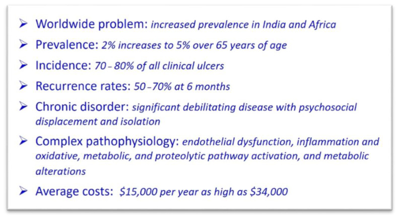
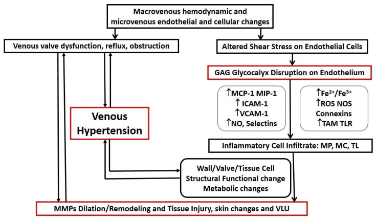
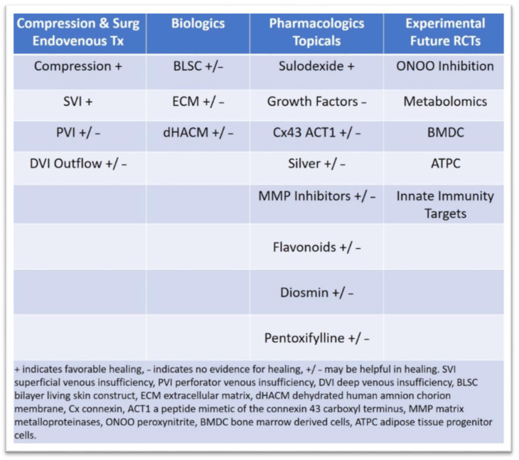
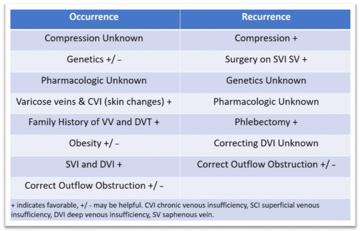
Venous leg ulcers (VLUs) are one of the most common ulcers of the lower extremity. VLU affects many individuals worldwide, could pose a significant socioeconomic burden to the healthcare system, and has major psychological and physical impacts on the affected individual. VLU often occurs in association with post-thrombotic syndrome, advanced chronic venous disease, varicose veins, and venous hypertension. Several demographic, genetic, and environmental factors could trigger chronic venous disease with venous dilation, incompetent valves, venous reflux, and venous hypertension. Endothelial cell injury and changes in the glycocalyx, venous shear-stress, and adhesion molecules could be initiating events in VLU. Increased endothelial cell permeability and leukocyte infiltration, and increases in inflammatory cytokines, matrix metalloproteinases (MMPs), reactive oxygen and nitrogen species, iron deposition, and tissue metabolites also contribute to the pathogenesis of VLU. Treatment of VLU includes compression therapy and endovenous ablation to occlude the axial reflux. Other interventional approaches such as subfascial endoscopic perforator surgery and iliac venous stent have shown mixed results. With good wound care and compression therapy, VLU usually heals within 6 months. VLU healing involves orchestrated processes including hemostasis, inflammation, proliferation, and remodeling and the contribution of different cells including leukocytes, platelets, fibroblasts, vascular smooth muscle cells, endothelial cells, and keratinocytes as well as the release of various biomolecules including transforming growth factor-β, cytokines, chemokines, MMPs, tissue inhibitors of MMPs (TIMPs), elastase, urokinase plasminogen activator, fibrin, collagen, and albumin. Alterations in any of these physiological wound closure processes could delay VLU healing. Also, these histological and soluble biomarkers can be used for VLU diagnosis and assessment of its progression, responsiveness to healing, and prognosis. If not treated adequately, VLU could progress to non-healed or granulating VLU, causing physical immobility, reduced quality of life, cellulitis, severe infections, osteomyelitis, and neoplastic transformation. Recalcitrant VLU shows prolonged healing time with advanced age, obesity, nutritional deficiencies, colder temperature, preexisting venous disease, deep venous thrombosis, and larger wound area. VLU also has a high, 50-70% recurrence rate, likely due to noncompliance with compression therapy, failure of surgical procedures, incorrect ulcer diagnosis, progression of venous disease, and poorly understood pathophysiology. Understanding the molecular pathways underlying VLU has led to new lines of therapy with significant promise including biologics such as bilayer living skin construct, fibroblast derivatives, and extracellular matrices and non-biologic products such as poly-N-acetyl glucosamine, human placental membranes amnion/chorion allografts, ACT1 peptide inhibitor of connexin 43, sulodexide, growth factors, silver dressings, MMP inhibitors, and modulators of reactive oxygen and nitrogen species, the immune response and tissue metabolites. Preventive measures including compression therapy and venotonics could also reduce the risk of progression to chronic venous insufficiency and VLU in susceptible individuals.
Keywords: biochemistry; chronic venous disease; chronic venous insufficiency; clinical medicine; healing; pathophysiology; therapy; venous leg ulcer.
Conflict of interest statement
The authors declare no conflict of interest. This review was written independently. The authors have given talks, attended conferences, and participated in advisory boards and trials sponsored by various pharmaceutical companies that had no role in the design of the review, in the interpretation of data and in the writing of the manuscript, or in the decision to publish the results.
Figures
References
-
- O’Donnell T.F., Jr., Passman M.A., Marston W.A., Ennis W.J., Dalsing M., Kistner R.L., Lurie F., Henke P.K., Gloviczki M.L., Eklöf B.G., et al. Management of venous leg ulcers: Clinical practice guidelines of the Society for Vascular Surgery® and the American Venous Forum. J. Vasc. Surg. 2014;60:3S–59S. doi: 10.1016/j.jvs.2014.04.049. - DOI - PubMed
Publication types
LinkOut - more resources
Full Text Sources
Medical
