

How to Build and to Protect the Neuromuscular Junction: The Role of the Glial Cell Line-Derived Neurotrophic Factor
- PMID: 33374485
- PMCID: PMC7794999
- DOI: 10.3390/ijms22010136
How to Build and to Protect the Neuromuscular Junction: The Role of the Glial Cell Line-Derived Neurotrophic Factor
Abstract
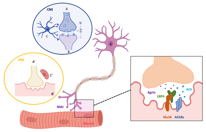
The neuromuscular junction (NMJ) is at the crossroad between the nervous system (NS) and the muscle. Following neurotransmitter release from the motor neurons (MNs), muscle contraction occurs and movement is generated. Besides eliciting muscle contraction, the NMJ represents a site of chemical bidirectional interplay between nerve and muscle with the active participation of Schwann cells. Indeed, signals originating from the muscle play an important role in synapse formation, stabilization, maintenance and function, both in development and adulthood. We focus here on the contribution of the Glial cell line-Derived Neurotrophic Factor (GDNF) to these processes and to its potential role in the protection of the NMJ during neurodegeneration. Historically related to the maintenance and survival of dopaminergic neurons of the substantia nigra, GDNF also plays a fundamental role in the peripheral NS (PNS). At this level, it promotes muscle trophism and it participates to the functionality of synapses. Moreover, compared to the other neurotrophic factors, GDNF shows unique peculiarities, which make its contribution essential in neurodegenerative disorders. While describing the known structural and functional changes occurring at the NMJ during neurodegeneration, we highlight the role of GDNF in the NMJ-muscle cross-talk and we review its therapeutic potential in counteracting the degenerative process occurring in the PNS in progressive and severe diseases such as Alzheimer's disease (AD), Amyotrophic Lateral Sclerosis (ALS) and Spinal Muscular Atrophy (SMA). We also describe functional 3D neuromuscular co-culture systems that have been recently developed as a model for studying both NMJ formation in vitro and its involvement in neuromuscular disorders.
Keywords: 3D neuromuscular model; AD; ALS; GDNF; SMA; motor neuron diseases; neurodegenerative diseases; neuromuscular junctions; neurotrophic factors.
Conflict of interest statement
The authors declare that the research was conducted in the absence of any commercial or financial relationships that could be construed as a potential conflict of interest.
Figures

References
-
- Castillo X., Castro-Obregón S., Gutiérrez-Becker B., Gutiérrez-Ospina G., Karalis N., Khalil A.A., Lopez-Noguerola J.S., Rodríguez L.L., Martínez-Martínez E., Perez-Cruz C., et al. Re-thinking the Etiological Framework of Neurodegeneration. Front. Neurosci. 2019;13:728. doi: 10.3389/fnins.2019.00728. - DOI - PMC - PubMed
Publication types
MeSH terms
Substances
Grants and funding
LinkOut - more resources
Full Text Sources
Miscellaneous
