Review
. 2020 Dec 23;10(1):3.
doi: 10.3390/antibiotics10010003.

Biofilms as Promoters of Bacterial Antibiotic Resistance and Tolerance

Affiliations
Review

Biofilms as Promoters of Bacterial Antibiotic Resistance and Tolerance

Cristina Uruén et al. Antibiotics (Basel). .

Abstract

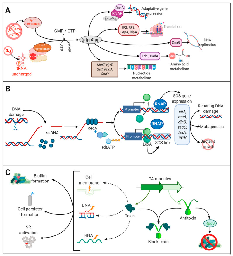
Multidrug resistant bacteria are a global threat for human and animal health. However, they are only part of the problem of antibiotic failure. Another bacterial strategy that contributes to their capacity to withstand antimicrobials is the formation of biofilms. Biofilms are associations of microorganisms embedded a self-produced extracellular matrix. They create particular environments that confer bacterial tolerance and resistance to antibiotics by different mechanisms that depend upon factors such as biofilm composition, architecture, the stage of biofilm development, and growth conditions. The biofilm structure hinders the penetration of antibiotics and may prevent the accumulation of bactericidal concentrations throughout the entire biofilm. In addition, gradients of dispersion of nutrients and oxygen within the biofilm generate different metabolic states of individual cells and favor the development of antibiotic tolerance and bacterial persistence. Furthermore, antimicrobial resistance may develop within biofilms through a variety of mechanisms. The expression of efflux pumps may be induced in various parts of the biofilm and the mutation frequency is induced, while the presence of extracellular DNA and the close contact between cells favor horizontal gene transfer. A deep understanding of the mechanisms by which biofilms cause tolerance/resistance to antibiotics helps to develop novel strategies to fight these infections.

Keywords: antibiotic resistance; antibiotic tolerance; biofilm control; biofilms; multidrug-resistant bacteria; recalcitrance.


Conflict of interest statement

The authors declare no conflict of interest.

Figures

Figure 1
Figure 1
Variable architecture of biofilms. (A) Biofilms of five species (Salmonella enterica, Escherichia coli, Pseudomonas aeruginosa Staphylococcus aureus, Enterococcus faecalis) were formed under static conditions on abiotic surfaces during 24 h and were stained with Syto9, a green fluorescent nucleic acid marker. Reprinted from [51] with permission from Elsevier. (B) Strains of Neisseria meningitidis HB-1 and α153 and derivatives, which do or do not produce the autotransporters AutA and AutB (as indicated), formed biofilms under flow conditions during 14 h and were stained with the LIVE/DEAD Backlight bacterial viability stain (where red cells are dead and green cells are live). Reproduced from [52,53].
Figure 2
Figure 2
Mechanisms of biofilms recalcitrance. Biofilm recalcitrance comprises a combination of antibiotic (ATB) resistance mechanisms (right panel) and ATB tolerance mechanisms (left panel). Resistance mechanisms confer the ability to survive and grow at increased ATB concentrations for long periods and involve horizontal genetic transfer (HGT), hypermutation, and quorum sensing (QS), leading to transport of antibiotics via efflux pumps, reduced permeability of the outer membrane, or production of enzymes that inactivate ATB. The type of AMR (Box 1) is indicated in green. In contrast, tolerance mechanisms lead microorganisms to survive at increased ATB concentrations temporally, and involve activation of stress responses (SOS response, stringent response SR)) and hypoxia, leading to activation of a quiescent state, anaerobic metabolism, decrease of membrane potential, and moderate increase in efflux pump expression. Arrowhead lines indicate the interrelation between mechanisms. CM: cytoplasmic membrane.
Figure 3
Figure 3
The stringent response, toxin-antitoxin, and SOS response pathways. (A) The stringent response (SR) is triggered by several stress conditions (amino-acid, carbon and iron deprivation or membrane damage) that activate the production (p)ppGpp by the synthetases RelA and SpoT and homologues. (p)ppGpp reprograms cell metabolism through the interaction with proteins involved in translation, transcription, replication, amino-acid metabolism, and nucleotide metabolism. (B) The SOS response is triggered by damaged DNA. Single-stranded (ssDNA) is detected by RecA. In the presence of (d)ATP, RecA is activated causing self-cleavage of LexA. LexA is a dimer that represses the transcription of SOS genes, which harbor an SOS box in their promoter. Cleavage of LexA leads to activation of the SOS genes inducing a repertoire of activities as indicated. The lexA gene also contains an SOS box; therefore, LexA production is self-regulated. Once the DNA is repaired, LexA represses the SOS response. (C) Several toxin-antitoxin (TA) modules are dispersed on the chromosome and are further classified according to the nature of the antitoxin and its mechanism of action. They are constituted by two genes, one encoding a toxin, with specific activities against target molecules (DNA, RNA, membrane, cell wall synthesis, ATP), and the other an antitoxin that binds to the toxin and inhibits its toxic activity. Under normal conditions, toxin and antitoxin are equally produced and thus the toxin does not exert its function. However, under stress conditions, the antitoxin can be degraded, and the toxin is free for toxic reactions. SR and SOS responses increase the production of ClpXP and Lon proteases, which activates the toxin by degradation of the antitoxin TA modules production. The toxic activity has several repercussions on cell biology.

References

    1. O’Neill J. Tackling Drug-Resistant Infections Globally: Final Report and Recommendations; the Review on Antimicrobial Resistance. [(accessed on 5 August 2020)];2016 Available online: https://amr-review.org/sites/default/files/160525_Final%20paper_with%20c....
    1. WHO (World Health Organization) High Levels of Antibiotic Resistance Found Worldwide, New Data Shows. [(accessed on 8 August 2020)];2018 Available online: https://www.who.int/news-room/detail/29-01-2018-high-levels-of-antibioti....
    1. EFSA (European Food Safety Authority) ECDC (European Centre for Disease Prevention and Control) The European Union summary report on antimicrobial resistance in zoonotic and indicator bacteria from humans, animals and food in 2017/2018. EFSA J. 2020;18:e06007. doi: 10.2903/j.efsa.2020.6007. - DOI - PMC - PubMed
    1. Collignon P. Fluoroquinolone use in food animals. Emerg. Infect. Dis. 2005;11:1789–1790. doi: 10.3201/eid1111.040630. - DOI - PMC - PubMed
    1. Landers T.F., Cohen B., Wittum T.E., Larson E.L. A review of antibiotic use in food animals: Perspective, policy, and potential. Public Health Rep. 2012;127:4–22. doi: 10.1177/003335491212700103. - DOI - PMC - PubMed

LinkOut - more resources