Review
. 2020 Dec 23;22(1):73.
doi: 10.3390/ijms22010073.

The Many Faces of CD4+ T Cells: Immunological and Structural Characteristics

Affiliations
Review

The Many Faces of CD4+ T Cells: Immunological and Structural Characteristics

Demetra S M Chatzileontiadou et al. Int J Mol Sci. .

Abstract

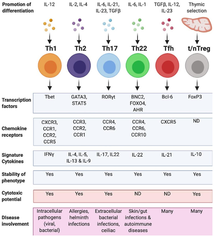
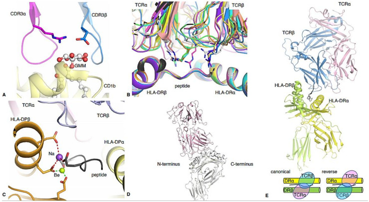
As a major arm of the cellular immune response, CD4+ T cells are important in the control and clearance of infections. Primarily described as helpers, CD4+ T cells play an integral role in the development and activation of B cells and CD8+ T cells. CD4+ T cells are incredibly heterogeneous, and can be divided into six main lineages based on distinct profiles, namely T helper 1, 2, 17 and 22 (Th1, Th2, Th17, Th22), regulatory T cells (Treg) and T follicular helper cells (Tfh). Recent advances in structural biology have allowed for a detailed characterisation of the molecular mechanisms that drive CD4+ T cell recognition. In this review, we discuss the defining features of the main human CD4+ T cell lineages and their role in immunity, as well as their structural characteristics underlying their detection of pathogens.

Keywords: CD4 T cell; Tfh; Th1; Th17; Th2; Th22; Treg; human leukocyte antigen (HLA); structural biology.


Conflict of interest statement

The authors declare no conflict of interest.

Figures

Figure 1
Figure 1
Structural insights into human CD4 TCR recognition. (A) Overview of the pHLA-II antigen binding cleft from the top and (B) side. The HLA-II α-chain is in orange and the β-chain is in green. The peptide, in black, is bound in the open cleft of HLA-II, with potential overhang residues at each end of the cleft. (C) Structure of a CD4 TCR-pHLA-II complex. The F24 TCR is in pink (α-chain) and blue (β-chain), and binding atop the HLA-DR11 (β-chain in orange, HLA-DRα in green) presenting the RQ13 HIV peptide (black). (D) Overlay of all known human CD4 TCR-pHLA-II complexes (except GEM42 TCR-CD1b-GMM), further detailed in Table 1.
Figure 2
Figure 2
Summary of human CD4+ T cell subsets. CD4+ T cell helper subsets and their characteristics. Each T helper subtype (Th1, Th2, Th17, Th22, Tfh and thymic/natural (t/n) Treg) is associated with different differentiation agonists, the expression of different transcription factors, chemokine receptors and the secretion of specific cytokines. A CD4+ T cell subset also displays a stable phenotype and may have cytotoxic potential. Some subsets have not been defined as having certain characteristics (ND). Each of these characteristics contributes to the cell’s ability to respond in a range of different diseases. Figure created using Biorender.com.
Figure 3
Figure 3
Timeline for the discovery of CD4+ T cell subsets. Timeline of CD4+ T cell subset discovery and major pHLA-TCR structures. Blue timepoints indicate major immunological discoveries in the identification of the CD4+ T cell subsets and red timepoints indicate the publication of major pHLA-TCR structures. The disease from which the peptide was derived or the type of helper subset of the TCR is written in parentheses. Figure created using Biorender.com.
Figure 4
Figure 4
Structural highlights of some human CD4 TCR-pHLA-II complexes. (A) GEM42 TCR CDR3α loop in pink, and CDR3β loop in blue, wrapping the GMM lipid-based antigen (white stick and spheres) presented by the HLA-like molecule CD1b (yellow). (B) Structural overlay of all gluten-specific TCRs bound to their antigen, alignment made on the antigen binding cleft. The molecules are represented as cartoons, and the characteristic Arginine residue from TCRs is shown as a stick. The complexes are coloured in green (PDB code: 4Z7U), cyan (PDB code: 4Z7V), pink (PDB code: 4Z7W), yellow (PDB code: 5K9S), salmon (PDB code: 5KSA), white (PDB code: 5KSB), purple (PDB code: 4OZH), pale yellow (PDB code: 4OZG), blue (PDB code: 4OZF) and orange (PDB code: 4GG6). (C) AV22 TCR (α-chain in pink and β-chain in blue) binding to the HLA-DP2 molecule (α-chain in yellow and β-chain in orange) complexed with the mimotope M2 peptide (grey) and Be (green sphere) and Na (purple sphere). The bonds formed between the pHLA and the Na or Be are represented by red or green dashed lines, respectively. (D) Representative docking topology of the majority of CD4 Th17 TCRs binding towards the N-terminus of the pHLA, represented by the 3A6 TCR in light pink [29]. (E) CD4 Treg FS18 TCR in complex with the proinsulin-HLA-DR4 (HLA-DRβ chain in green, HLA-DRα in yellow, peptide in black). The TCRα chain is in pink and over the HLA-DRα chain, while the TCRβ chain is coloured in blue and docks over the HLA-DRβ chain, exhibiting a reversed topology. The bottom panel show a schematic of canonical and reversed docking topology for αβTCRs, respectively.

References

    1. Rossjohn J., Gras S., Miles J.J., Turner S.J., Godfrey D.I., McCluskey J. T cell antigen receptor recognition of antigen-presenting molecules. Annu. Rev. Immunol. 2015;33:169–200. doi: 10.1146/annurev-immunol-032414-112334. - DOI - PubMed
    1. Ohno Y., Kitamura H., Takahashi N., Ohtake J., Kaneumi S., Sumida K., Homma S., Kawamura H., Minagawa N., Shibasaki S., et al. IL-6 down-regulates HLA class II expression and IL-12 production of human dendritic cells to impair activation of antigen-specific CD4(+) T cells. Cancer Immunol. Immunother. 2016;65:193–204. doi: 10.1007/s00262-015-1791-4. - DOI - PMC - PubMed
    1. Rijvers L., Melief M.-J., Van Langelaar J., Vries R.M.V.D.V.D., Wierenga-Wolf A.F., Koetzier S.C., Priatel J., Jorritsma T., Van Ham S.M., Hintzen R.Q., et al. The Role of Autoimmunity-Related Gene CLEC16A in the B Cell Receptor-Mediated HLA Class II Pathway. J. Immunol. 2020;205:945–956. doi: 10.4049/jimmunol.1901409. - DOI - PubMed
    1. Codolo G., Toffoletto M., Chemello F., Coletta S., Teixidor G.S., Battaggia G., Munari G., Fassan M., Cagnin S., De Bernard M. Helicobacter pylori Dampens HLA-II Expression on Macrophages via the Up-Regulation of miRNAs Targeting CIITA. Front. Immunol. 2020;10:2923. doi: 10.3389/fimmu.2019.02923. - DOI - PMC - PubMed
    1. Wosen J.E., Mukhopadhyay D., Macaubas C., Mellins E.D. Epithelial MHC Class II Expression and Its Role in Antigen Presentation in the Gastrointestinal and Respiratory Tracts. Front. Immunol. 2018;9:2144. doi: 10.3389/fimmu.2018.02144. - DOI - PMC - PubMed

MeSH terms

Substances