. 2020 Dec 22;12(1):3.
doi: 10.3390/genes12010003.

Oxidative Stress, Glutathione Metabolism, and Liver Regeneration Pathways Are Activated in Hereditary Tyrosinemia Type 1 Mice upon Short-Term Nitisinone Discontinuation

Affiliations

Oxidative Stress, Glutathione Metabolism, and Liver Regeneration Pathways Are Activated in Hereditary Tyrosinemia Type 1 Mice upon Short-Term Nitisinone Discontinuation

Haaike Colemonts-Vroninks et al. Genes (Basel). .

Abstract

Hereditary tyrosinemia type 1 (HT1) is an inherited condition in which the body is unable to break down the amino acid tyrosine due to mutations in the fumarylacetoacetate hydrolase (FAH) gene, coding for the final enzyme of the tyrosine degradation pathway. As a consequence, HT1 patients accumulate toxic tyrosine derivatives causing severe liver damage. Since its introduction, the drug nitisinone (NTBC) has offered a life-saving treatment that inhibits the upstream enzyme 4-hydroxyphenylpyruvate dioxygenase (HPD), thereby preventing production of downstream toxic metabolites. However, HT1 patients under NTBC therapy remain unable to degrade tyrosine. To control the disease and side-effects of the drug, HT1 patients need to take NTBC as an adjunct to a lifelong tyrosine and phenylalanine restricted diet. As a consequence of this strict therapeutic regime, drug compliance issues can arise with significant influence on patient health. In this study, we investigated the molecular impact of short-term NTBC therapy discontinuation on liver tissue of Fah-deficient mice. We found that after seven days of NTBC withdrawal, molecular pathways related to oxidative stress, glutathione metabolism, and liver regeneration were mostly affected. More specifically, NRF2-mediated oxidative stress response and several toxicological gene classes related to reactive oxygen species metabolism were significantly modulated. We observed that the expression of several key glutathione metabolism related genes including Slc7a11 and Ggt1 was highly increased after short-term NTBC therapy deprivation. This stress response was associated with the transcriptional activation of several markers of liver progenitor cells including Atf3, Cyr61, Ddr1, Epcam, Elovl7, and Glis3, indicating a concreted activation of liver regeneration early after NTBC withdrawal.

Keywords: glutathione metabolism; hereditary liver disease; liver regeneration; nitisinone; oxidative stress; transcriptomics; tyrosinemia type 1.


Conflict of interest statement

The authors declare that they have no conflict of interest.

Figures

Figure 1
Figure 1
Tyrosine degradation pathway in liver cells. Loss of function of the fumarylacetoacetate hydrolase (FAH) enzyme causes the accumulation of toxic intermediate tyrosine metabolites maleyl- and fumarylacetoacetate, and subsequently, the production of succinylacetone through an alternative metabolization route. Nitisinone (NTBC) is a potent inhibitor of the upstream hydroxyphenylpyruvate dioxygenase (HPD) enzyme that prevents the formation of these toxic metabolites by providing a therapeutic block. Abbreviations: PAH, phenylalanine hydroxylase; TAT, tyrosine aminotransferase; HPD, 4- hydroxyphenylpyruvate dioxygenase; HGD, homogentisate dioxygenase; MAI, maleylacetoacetate isomerase; FAH, fumarylacetoacetate hydrolase, NTBC, nitisinone.
Figure 2
Figure 2
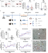
Molecular and biochemical evaluation of the FRG mouse model. FRG mouse livers are characterized by (A) the absence of Fah and (B) the presence of Hpd protein expression using western blot with healthy wildtype (WT) C57Bl/6 mice as positive controls. (C) Schematic representation of the NTBC drug therapy discontinuation experiment in FRG mice and subsequent sample collection. (D,E) Gene expression analysis of mouse albumin (Alb) and alpha-fetoprotein (Afp) by RT-qPCR. (F) Quantification of NTBC blood levels and daily follow up of (G) succinylacetone, (H) L-tyrosine and (I) L-phenylalanine levels using LC-MS/MS analyses of dried blood spots (DBS). Histopathological analysis of FRG mouse livers under (J) continuous NTBC therapy and (K) seven days post NTBC withdrawal by hematoxylin staining showing large dysmorphic nuclei (N), oval-like cells (black arrow) and small lymphoid cells (yellow arrow). * significantly increased; ** significantly decreased using a two-tailed Mann–Whitney test (p < 0.05).
Figure 3
Figure 3
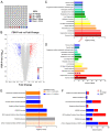
Whole transcriptome analyses of FRG liver tissue after seven days of NTBC therapy discontinuation versus continuous treatment. (A) Percentage of modulated genes in liver tissue. (B) Volcano plot displaying 5-fold up and down regulated genes with FDR p-value ≤ 0.05. (C,D) Toxicological gene class grouping of 5-fold up and down regulated genes showing the Benjamini–Hochberg (B–H) p-value of overlap and amount of modulated genes. (E,F) Canonical pathway analyses showing B–H p-value of overlap, activation z-scores and amount of modulated genes.
Figure 4
Figure 4
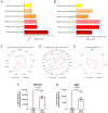
NTBC therapy discontinuation activates oxidative stress responses, reactive oxygen species (ROS) formation and glutathione metabolism. (A,B) ROS-related gene class grouping of 5-fold up and down regulated genes showing B–H p-value of overlap and amount of modulated genes. Spider graphs showing differentially-expressed genes related to (C) Nrf2-mediated oxidative stress response, (D) reactive oxygen species formation, and (E) glutathione metabolism. (F,G) Gene expression analysis of top increased ROS-related genes by RT-qPCR. * significantly increased using a two-tailed Mann–Whitney test (p < 0.05).
Figure 5
Figure 5
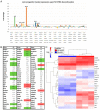
Liver progenitor cell (LPC)/biliary epithelial cell (BEC) marker expression in Fah-deficient mouse livers after short-term NTBC therapy discontinuation. (A,B) Gene expression profiling of LPC/BEC markers using microarray analyses. (C) Hierarchical clustering of differentially-expressed LPC/BEC markers.
Figure 6
Figure 6
Gene expression analysis of LPC/BEC identification markers by RT-qPCR. * significantly increased using a two-tailed Mann–Whitney test (p < 0.05).

References

    1. De Laet C., Dionisi-Vici C., Leonard J.V., McKiernan P., Mitchell G., Monti L., de Baulny H.O., Pintos-Morell G., Spiekerkötter U. Recommendations for the management of tyrosinaemia type 1. Orphanet J. Rare Dis. 2013;8:8. doi: 10.1186/1750-1172-8-8. - DOI - PMC - PubMed
    1. Alvarez F., Atkinson S., Bouchard M., Brunel-Guitton C., Buhas D., Bussières J.F., Dubois J., Fenyves D., Goodyer P., Gosselin M., et al. The Québec NTBC study. Adv. Exp. Med. Biol. 2017;959:187–195. - PubMed
    1. Larochelle J. Discovery of hereditary tyrosinemia in Saguenay-Lac St-Jean. Adv. Exp. Med. Biol. 2017;959:3–8. - PubMed
    1. Äärelä L., Hiltunen P., Soini T., Vuorela N., Huhtala H., Nevalainen P.I., Heikinheimo M., Kivelä L., Kurppa K. Type 1 tyrosinemia in Finland: A nationwide study. Orphanet J. Rare Dis. 2020;15:1–11. doi: 10.1186/s13023-020-01547-w. - DOI - PMC - PubMed
    1. Bliksrud Y.T., Brodtkorb E., Backe P.H., Woldseth B., Rootwelt H. Hereditary tyrosinaemia type 1 in Norway: Incidence and three novel small deletions in the fumarylacetoacetase gene. Scand. J. Clin. Lab. Invest. 2012;72:369–373. doi: 10.3109/00365513.2012.676210. - DOI - PubMed

Publication types

MeSH terms