Review
. 2020 Dec 22;11(1):2.
doi: 10.3390/biom11010002.

Molecules, Mechanisms, and Disorders of Self-Domestication: Keys for Understanding Emotional and Social Communication from an Evolutionary Perspective

Affiliations
Review

Molecules, Mechanisms, and Disorders of Self-Domestication: Keys for Understanding Emotional and Social Communication from an Evolutionary Perspective

Goran Šimić et al. Biomolecules. .

Abstract

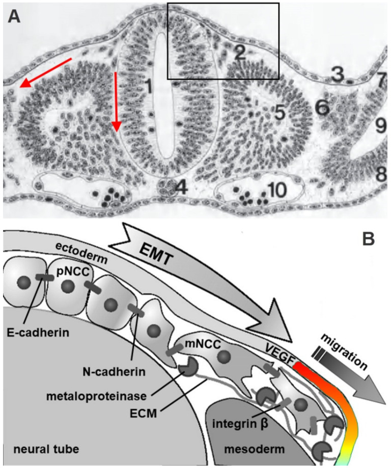
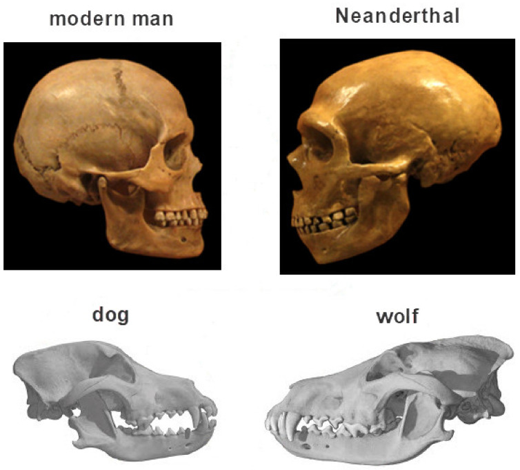
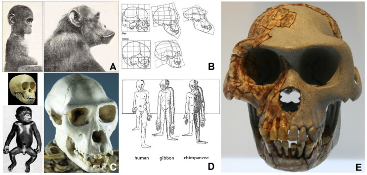
The neural crest hypothesis states that the phenotypic features of the domestication syndrome are due to a reduced number or disruption of neural crest cells (NCCs) migration, as these cells differentiate at their final destinations and proliferate into different tissues whose activity is reduced by domestication. Comparing the phenotypic characteristics of modern and prehistoric man, it is clear that during their recent evolutionary past, humans also went through a process of self-domestication with a simultaneous prolongation of the period of socialization. This has led to the development of social abilities and skills, especially language, as well as neoteny. Disorders of neural crest cell development and migration lead to many different conditions such as Waardenburg syndrome, Hirschsprung disease, fetal alcohol syndrome, DiGeorge and Treacher-Collins syndrome, for which the mechanisms are already relatively well-known. However, for others, such as Williams-Beuren syndrome and schizophrenia that have the characteristics of hyperdomestication, and autism spectrum disorders, and 7dupASD syndrome that have the characteristics of hypodomestication, much less is known. Thus, deciphering the biological determinants of disordered self-domestication has great potential for elucidating the normal and disturbed ontogenesis of humans, as well as for the understanding of evolution of mammals in general.

Keywords: chemoattractants; chemorepellents; epithelial-mesenchymal transition (EMT); extracellular matrix molecules; fibroblast growth factor (FGF); methyl-CpG-binding protein 2 (MeCP2); neural crest cells (NCCs); self-domestication; thyroid hormones; vascular endothelial growth factor (VEGF).


Conflict of interest statement

The authors declare no conflict of interest.

Figures

Figure 1
Figure 1
Schematic representation of neural tube development, neural crest cells (NCCs) migration, and some associations of NCCs with phenotypic traits of the domestication syndrome in mammals. The upper part of the schematics is made according to Marieb and Hoehn (2018) [27] and Kaltschmidt et al. (2018) [28]. The Waddington’s epigenetic landscape (“canalization”) on the right side illustrates contribution of epigenetic changes on NCCs migration, differentiation, and proliferation, and is made according to Strobl-Mazzulla and Bronner (2014) [29]. For the purpose of this schematic representation, no distinction is made between anterior (cephalic) and posterior somatic NCCs.
Figure 2
Figure 2
Delamination of NCCs from neuroectoderm via ectodermal-mesenchymal transition and migration of NCCs via ventrodorsal and ventromedial pathways. Cross-section through a human embryo on day 24 of gestation (crown-rump length 3 mm). The lower part of figure (B) represents enlarged inset from the upper part (A). Legend: 1, neural tube; 2, neural crest migratory cells; 3, ectoderm; 4, dorsal chord (notochord); 5, somite; 6, intermediate mesoderm; 7, somatopleura; 8, splanchnopleura; 9, intraembryonic whole; 10, aorta; a red medial (vertical) arrow between the neural tube and the somite indicates the ventromedial direction of NCCs migration, a red lateral arrow between the ectoderm and the somite indicates the dorsolateral direction of NCCs migration; ECM, extracellular matrix; EMT, epithelial-mesenchymal transition; mNCC, migratory neural crest cells; pNCC, premigratory neural crest cells, VEGF, vascular endothelial growth factor. The spatio-temporal expression pattern of VEGF in the ectoderm (colored) regulates the NCCs migratory front. The upper part of figure (A) is from Christ (1985) [32], and the lower part of the image is schematized according to Szabo and Mayor (2018) [31] and McLennan et al. (2010) [33].
Figure 3
Figure 3
Schematic representation of the dynamics of neural crest cell migration in the rostral and caudal part of the human embryo. Migration begins around day 22 and ends by day 31 of gestation. The total number of somites in human is 37 as out of the original 42–44, some of the most caudal ones rapidly disappear [36].
Figure 4
Figure 4
Simplified schematics of stepwise activation of the regulatory gene modules and networks for NCCs development, delamination, specification, migration, proliferation, and differentiation. Genes for specification of the first signaling module, which induces neural crest, do not exist in invertebrates. Scheme is modified according to Green et al. (2015) [39] and Simões-Costa et al. (2015) [35]. Today, it is known that the specification and migration modules include more genes, including Twist, Sox5, Sox9, Myc, Tfap2, Sox10, Myb, RxrG, Myc, and many others.
Figure 5
Figure 5
Comparison of dog domestication and human self-domestication. Illustration modified from Theofanopoulou et al. (2017) [45]. Photographs of the skull of modern man (Homo sapiens) and prehistoric man (Homo neandertalensis) are from commons.wikimedia.org. Similar to differences between dog and wolf, note that the skull of modern man is much more neotenic compared to the skull of a Neanderthal (smaller skull and brain, oval forehead, less protruded nasal bone, smaller teeth, orthognathia), which can be said to be gerontomorphic.
Figure 6
Figure 6
The development of the idea of neoteny in human. (A). Photograph of a chimpanzee child and an adult chimpanzee from Naef (1926) [60]. (B). The top row shows the growth and shape of the skull in a fetus, cub, and adult chimpanzee, and the bottom row shows the skull of a fetus and an adult human. The direction of transformation is the same: negative skull allometry is seen as the skull grow slower than the rest of the body and is therefore being proportionally smaller in adults than in children (hypoallometry) and positive allometry of the face and jaw growing faster than the rest of the body and therefore proportionally higher in adults than in children (hyperallometry); image from Starck and Kummer (1962) [61]. (C). Skulls of young and adult chimpanzees (from commons.wikimedia.org) for comparison with Figures A and B. In the lower left corner is a picture of a seven-month-old chimpanzee fetus. Hair growth can be seen on the head, in the eyebrow area, the edges of the eyelids, lips, and cheeks, i.e., in those places where we see them today in adults, which is considered as another proof of neoteny in human; image from the University of Wisconsin Public Digital Repository website (www.omnia.ie). (D). Comparative view of infant and adult human, gibbon, and chimpanzee, all aligned with height in sitting position (frame represents trunk and head size). Lesser apes (gibbons) have extremely long front limbs, even at birth. The legs always grow faster than the trunk, especially in a human. The newborn has relatively short arms at birth. Note smaller hands and feet in man. The image transferred from Verhulst (1993) [62], originally drawn by Schultz (1926) [63]. (E). Cast of a reconstructed skull of the Ardipithecus ramidus. Note the flattened face and pronounced orthognathia (the cast is on display at the State Archaeological Museum in Chemnitz, Federal Republic of Germany; image from commons.wikimedia.org).
Figure 7
Figure 7
Illustration of the formation of asymmetry of the cerebral hemispheres. Since it was first described by Paul Yakovlev, it is also called Yakovlev’s twisting (torque) of the brain in a counterclockwise direction (Yakovlev cerebral anticlockwise torque). In the diagram, the twist is not shown from above, but from below (so in this view it is clockwise). Twisting also leads to asymmetry of the protrusion of the inner surface of the skull bones (petalia) as the bones adapt to the shape of the brain (and not vice versa). The scheme is from commons.wikimedia.org.
Figure 8
Figure 8
Time-lapse of major discoveries in regard to genes associated with autism spectrum disorder (ASD). FXS, Fragile X syndrome; iPSC, induced pluripotent stem cells; TSC, tuberous sclerosis complex. See text for details.
Figure 9
Figure 9
Early excessive growth of head circumference in children with autism spectrum disorders (ASD). After reduced head circumference at birth, during the first year of life in children with ASD there is a pathologically increased growth in head circumference and the total size of the cerebellum and cerebellum. The graph is based on Courchesne et al. (2003) [196].
Figure 10
Figure 10
Schematic representation of increased serotonin levels in the blood (hyperserotonemia) in individuals with autism spectrum disorder (ASD). The blue curve presents the concentration of serotonin in the blood in control subjects. Graph based on Laboyer et al. (1999) [219].
Figure 11
Figure 11
Schematic representation of the five-point model of processes involved in the regulation of emotions according to Gross. For any emotional event, there are a number of steps on which emotion regulation can be applied. See text for details. Slightly modified from Gross (2013) [244].

Similar articles

Cited by

References

    1. Darwin C. The Variation of Plants and Animals under Domestication. John Murray; London, UK: 1868.
    1. Wilkins A.S., Wrangham R.W., Fitch W.T. The “domestication syndrome“ in mammals: A unified explanation based on neural crest cell behavior and genetics. Genetics. 2014;197:795–808. doi: 10.1534/genetics.114.165423. - DOI - PMC - PubMed
    1. Sánchez-Villagra M.R., Geiger M., Schneider R.A. The taming of the neural crest: A developmental perspective on the origins of morphological covariation in domesticated mammals. R. Soc. Open Sci. 2016;3:160107. doi: 10.1098/rsos.160107. - DOI - PMC - PubMed
    1. Wheat C.H., van der Bijl W., Wheat C.W. Morphology does not covary with predicted behavioral correlations of the domestication syndrome in dogs. BioRxiv. 2020 doi: 10.1101/660829. - DOI - PMC - PubMed
    1. Irving-Pease E.K., Ryan H., Jamieson A., Dimopoulos E.A. Paleogenomics of animal domestication. In: Lindqvist C., Rajora O., editors. Paleogenomics. Population Genomics. Springer; Berlin/Heidelberg, Germany: 2018. pp. 225–272.

Publication types

LinkOut - more resources