

Endothelium Infection and Dysregulation by SARS-CoV-2: Evidence and Caveats in COVID-19
- PMID: 33375371
- PMCID: PMC7823949
- DOI: 10.3390/v13010029
Endothelium Infection and Dysregulation by SARS-CoV-2: Evidence and Caveats in COVID-19
Abstract
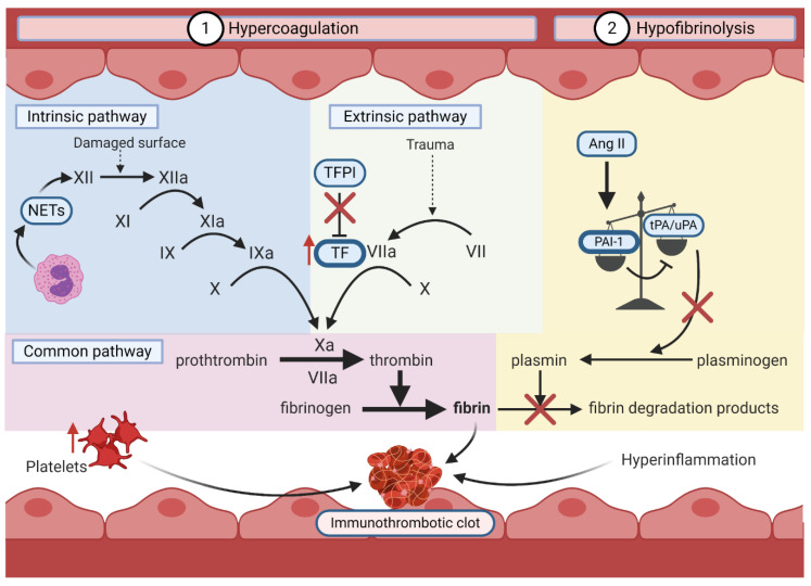
The ongoing pandemic of coronavirus disease 2019 (COVID-19) caused by the acute respiratory syndrome-coronavirus-2 (SARS-CoV-2) poses a persistent threat to global public health. Although primarily a respiratory illness, extrapulmonary manifestations of COVID-19 include gastrointestinal, cardiovascular, renal and neurological diseases. Recent studies suggest that dysfunction of the endothelium during COVID-19 may exacerbate these deleterious events by inciting inflammatory and microvascular thrombotic processes. Although controversial, there is evidence that SARS-CoV-2 may infect endothelial cells by binding to the angiotensin-converting enzyme 2 (ACE2) cellular receptor using the viral Spike protein. In this review, we explore current insights into the relationship between SARS-CoV-2 infection, endothelial dysfunction due to ACE2 downregulation, and deleterious pulmonary and extra-pulmonary immunothrombotic complications in severe COVID-19. We also discuss preclinical and clinical development of therapeutic agents targeting SARS-CoV-2-mediated endothelial dysfunction. Finally, we present evidence of SARS-CoV-2 replication in primary human lung and cardiac microvascular endothelial cells. Accordingly, in striving to understand the parameters that lead to severe disease in COVID-19 patients, it is important to consider how direct infection of endothelial cells by SARS-CoV-2 may contribute to this process.
Keywords: ACE2; ADAM17; COVID-19; RAAS; SARS-CoV-2; bradykinin–kallikrein pathway; endothelial dysfunction; immunothrombosis; pericyte; therapeutics.
Conflict of interest statement
The authors declare no conflict of interest.
Figures
Similar articles
-
SARS-CoV-2 Spike Protein Destabilizes Microvascular Homeostasis.Microbiol Spectr. 2021 Dec 22;9(3):e0073521. doi: 10.1128/Spectrum.00735-21. Epub 2021 Dec 22. Microbiol Spectr. 2021. PMID: 34935423 Free PMC article.
-
Kallikrein-kinin blockade in patients with COVID-19 to prevent acute respiratory distress syndrome.Elife. 2020 Apr 27;9:e57555. doi: 10.7554/eLife.57555. Elife. 2020. PMID: 32338605 Free PMC article.
-
COVID-19, Renin-Angiotensin System and Endothelial Dysfunction.Cells. 2020 Jul 9;9(7):1652. doi: 10.3390/cells9071652. Cells. 2020. PMID: 32660065 Free PMC article. Review.
-
SARS-CoV-2 cell entry receptor ACE2 mediated endothelial dysfunction leads to vascular thrombosis in COVID-19 patients.Med Hypotheses. 2020 Dec;145:110320. doi: 10.1016/j.mehy.2020.110320. Epub 2020 Sep 30. Med Hypotheses. 2020. PMID: 33032170 Free PMC article.
-
Pulmonary Endothelial Dysfunction and Thrombotic Complications in Patients with COVID-19.Am J Respir Cell Mol Biol. 2021 Apr;64(4):407-415. doi: 10.1165/rcmb.2020-0359PS. Am J Respir Cell Mol Biol. 2021. PMID: 33180562 Free PMC article. Review.
Cited by
-
Implications of SARS-Cov-2 infection on eNOS and iNOS activity: Consequences for the respiratory and vascular systems.Nitric Oxide. 2021 Jun 1;111-112:64-71. doi: 10.1016/j.niox.2021.04.003. Epub 2021 Apr 6. Nitric Oxide. 2021. PMID: 33831567 Free PMC article. Review.
-
Vascular dysfunction in COVID-19 patients: update on SARS-CoV-2 infection of endothelial cells and the role of long non-coding RNAs.Clin Sci (Lond). 2022 Nov 11;136(21):1571-1590. doi: 10.1042/CS20220235. Clin Sci (Lond). 2022. PMID: 36367091 Free PMC article.
-
Dexamethasone for Severe COVID-19: How Does It Work at Cellular and Molecular Levels?Int J Mol Sci. 2021 Jun 23;22(13):6764. doi: 10.3390/ijms22136764. Int J Mol Sci. 2021. PMID: 34201797 Free PMC article. Review.
-
Effect of SARS-CoV-2 Infection and BNT162b2 Vaccination on the mRNA Expression of Genes Associated with Angiogenesis.Int J Mol Sci. 2023 Nov 8;24(22):16094. doi: 10.3390/ijms242216094. Int J Mol Sci. 2023. PMID: 38003287 Free PMC article.
-
A survey on the safety of the SARS-CoV-2 vaccine among a population with stroke risk in China.Front Med (Lausanne). 2022 Sep 21;9:859682. doi: 10.3389/fmed.2022.859682. eCollection 2022. Front Med (Lausanne). 2022. PMID: 36213663 Free PMC article.
References
-
- Hui D.S., Azhar E.I., Madani T.A., Ntoumi F., Kock R., Dar O., Ippolito G., Mchugh T.D., Memish Z.A., Drosten C., et al. The continuing 2019-nCoV epidemic threat of novel coronaviruses to global health—The latest 2019 novel coronavirus outbreak in Wuhan, China. Int. J. Infect. Dis. 2020;91:264–266. doi: 10.1016/j.ijid.2020.01.009. - DOI - PMC - PubMed
-
- WHO Director-General’s Opening Remarks at the Media Briefing on COVID-19—11 March 2020. [(accessed on 28 June 2020)]; Available online: https://www.who.int/dg/speeches/detail/who-director-general-s-opening-re....
Publication types
MeSH terms
Substances
Grants and funding
LinkOut - more resources
Full Text Sources
Medical
Miscellaneous
