Review
. 2020 Dec 27;22(1):186.
doi: 10.3390/ijms22010186.

AMP-Activated Protein Kinase: Do We Need Activators or Inhibitors to Treat or Prevent Cancer?

Affiliations
Review

AMP-Activated Protein Kinase: Do We Need Activators or Inhibitors to Treat or Prevent Cancer?

Fiona M Russell et al. Int J Mol Sci. .

Abstract

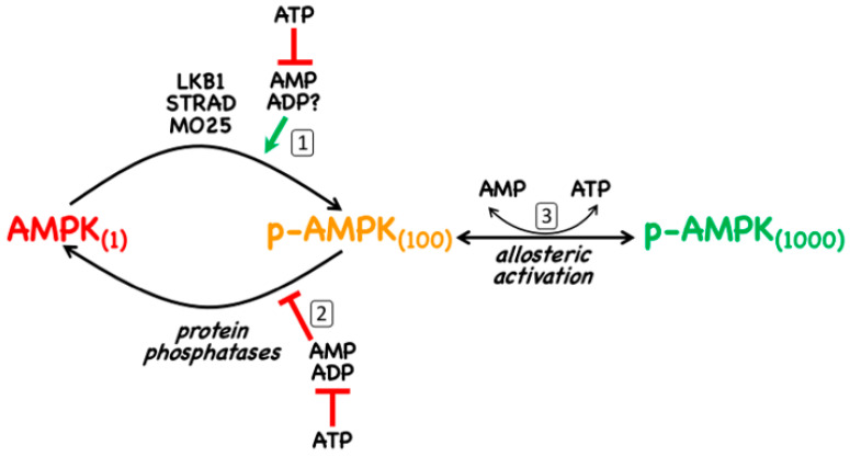
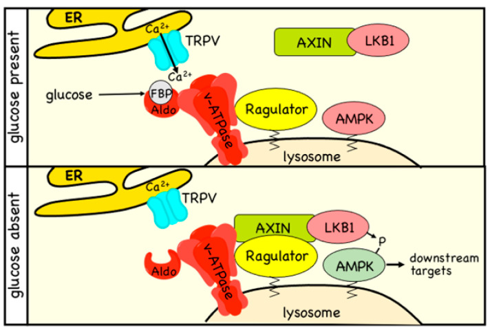
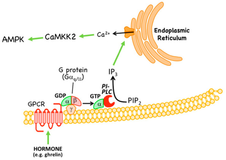
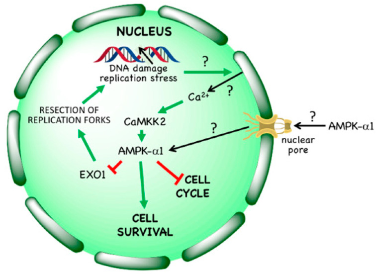
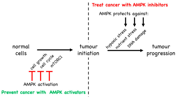
AMP-activated protein kinase (AMPK) is a key regulator of cellular energy balance. In response to metabolic stress, it acts to redress energy imbalance through promotion of ATP-generating catabolic processes and inhibition of ATP-consuming processes, including cell growth and proliferation. While findings that AMPK was a downstream effector of the tumour suppressor LKB1 indicated that it might act to repress tumourigenesis, more recent evidence suggests that AMPK can either suppress or promote cancer, depending on the context. Prior to tumourigenesis AMPK may indeed restrain aberrant growth, but once a cancer has arisen, AMPK may instead support survival of the cancer cells by adjusting their rate of growth to match their energy supply, as well as promoting genome stability. The two isoforms of the AMPK catalytic subunit may have distinct functions in human cancers, with the AMPK-α1 gene often being amplified, while the AMPK-α2 gene is more often mutated. The prevalence of metabolic disorders, such as obesity and Type 2 diabetes, has led to the development of a wide range of AMPK-activating drugs. While these might be useful as preventative therapeutics in individuals predisposed to cancer, it seems more likely that AMPK inhibitors, whose development has lagged behind that of activators, would be efficacious for the treatment of pre-existing cancers.

Keywords: AMP-activated protein kinase; AMPK; CaMKK2; LKB1; biguanides; cancer; kinase activators; kinase inhibitors; tumour promoters; tumour suppressors.


Conflict of interest statement

The authors declare no conflict of interest.

Figures

Figure 1
Figure 1
Domain maps of the three subunits of the AMPK heterotrimer: α, β and γ. Each subunit occurs as multiple isoforms. “Myr-N” refers to the myristoylated N-terminus of the β subunits. Other abbreviations/acronyms are defined in the text.
Figure 2
Figure 2
The canonical mechanism of activation of AMPK by adenine nucleotides. The action of adenine nucleotides on AMPK activity is threefold: (1) AMPK not phosphorylated on Thr172 (left) is phosphorylated by the LKB1:STRAD:MO25 complex and this reaction, which causes at least 100-fold activation, is promoted by the binding of AMP (and perhaps ADP) to AMPK itself; (2) binding of AMP or ADP to AMPK also causes a conformational change that inhibits dephosphorylation of Thr172 by protein phosphatases; (3) binding of AMP (but not ADP) causes up to 10-fold allosteric activation of AMPK. All three effects of AMP and/or ADP are antagonized by the binding of ATP. The figures in parentheses next to each form of AMPK are an indication of their approximate relative kinase activity.
Figure 3
Figure 3
Non-canonical mechanism of activation of AMPK by glucose starvation. The model is based on that in Li et al. [60]. In the presence of high glucose (top panel), a large proportion of aldolase (Aldo), which is bound to the v-ATPase proton pump on the lysosome, will have its substrate, the glycolytic intermediate fructose-1,6-bisphosphatase (FBP), bound to it. This causes the opening of TRPV Ca2+ channels located at ER:lysosome contact sites, maintaining the function of the v-ATPase and promoting mTORC1 activity via the Ragulator complex, which is anchored on the surface of the lysosome via lipid modifications. When glucose is absent or limiting (lower panel), aldolase will not be fully occupied by FBP, and it interacts with and inhibits the neighbouring TRPV channels. The v-ATPase is now inhibited, allowing an interaction between the Ragulator and the cytoplasmic Axin:LKB1 complex, and bringing LKB1 into the proximity of AMPK, which is then phosphorylated and activated. In this version of the model, a pool of AMPK is viewed as being permanently located at the lysosome due to the N-terminal myristoylation of the β subunit.
Figure 4
Figure 4
Non-canonical mechanism of activation of AMPK by hormones that trigger intracellular Ca2+ release. Hormones such as ghrelin bind to a G protein-coupled receptor (GPCR) that is coupled via G proteins containing Gαq/11 to activation of phosphatidylinositol-specific phospholipase C (PI-PLC). This triggers release of inositol-1,4,5-trisphosphate (IP3) from the plasma membrane phospholipid phosphatidylinositol-4,5-bisphosphate (PIP2). IP3 diffuses to the endoplasmic reticulum where it binds to IP3 receptors, triggering release of Ca2+ ions. These in turn activate Ca2+/calmodulin-dependent kinase kinase-2 (CaMKK2) which phosphorylates AMPK at Thr172, causing its activation.
Figure 5
Figure 5
Non-canonical mechanism of activation of AMPK in the cell nucleus by treatments that cause DNA damage. Agents such as ionizing radiation, cytotoxic drugs or DNA synthesis inhibitors cause DNA damage, either directly or following replicative stress. This causes release of Ca2+ ions within the nucleus, which activates CaMKK2 within that compartment. AMPK complexes containing AMPK-α1 also appear to translocate from the cytoplasm to the nucleus, where they are activated by CAMKK2-dependent phosphorylation at Thr172. AMPK activation enhances cell survival; it causes a G1 phase cell cycle arrest, and triggers phosphorylation of EXO1, limiting its ability to cause excessive resection at stalled replication forks. As indicated by question marks, several aspects of this mechanism remain unresolved: (i) how is the DNA damage sensed? (ii) what is the source of Ca2+ and its mechanism of release into the nucleus? and (iii) how is the translocation of AMPK-α1 to the nucleus achieved and regulated? The source of nuclear Ca2+ release is shown here as the nuclear envelope, but this is also unconfirmed.
Figure 6
Figure 6
Activation of AMPK by pro-drugs that are taken up into cells and converted into AMP analogues. TOP: AICA riboside is an adenosine analogue that is taken up into cells via adenosine transporters and converted by adenosine kinase into the equivalent nucleotide, ZMP, an AMP analogue that mimics the effects of AMP on the AMPK system. MIDDLE: C2 is a phosphonate analogue of AMP that is administered in an esterified form (C13) that diffuses across the plasma membrane and is converted by cellular esterases into C2. BOTTOM: cordycepin is an adenosine analogue that is taken up into cells via adenosine transporters and converted by adenosine kinase into the equivalent nucleotide, cordycepin-5′-monophosphate, an AMP analogue that mimics effects of AMP on the AMPK system.
Figure 7
Figure 7
Indirect activation of AMPK by drugs that interfere with cellular ATP synthesis. Depending on the cell type, drugs that inhibit glycolysis (e.g., 2-deoxyglucose) or that inhibit mitochondrial oxidative phosphorylation (especially Complex I inhibitors such as metformin, phenformin and berberine) cause an accumulation of ADP relative to ATP. This in turn causes displacement of the adenylate kinase reaction towards AMP and ATP, increasing AMP dramatically while having much smaller effects on ADP and ATP levels. The increase in AMP (and perhaps also ADP) relative to ATP then activates AMPK by the canonical mechanism shown in Figure 2.
Figure 8
Figure 8
Natural and pharmacological activators of AMPK that bind in the ADaM site. TOP: palmitoyl-CoA (and other long chain saturated and monounsaturated acyl-CoA esters) are believed to the natural ligands that bind at the ADaM site, but they only activate β1-containing complexes. MIDDLE: synthetic activators that are selective for β1-containing complexes. BOTTOM: synthetic pan-β activators that activate both β1- and β2-containing complexes.
Figure 9
Figure 9
The central dichotomy in the functions of AMPK as a target in cancer therapy. LEFT: by inhibiting biosynthesis and thus cell growth, the cell cycle and thus cell proliferation, and mTORC1 and thus metabolic changes conducive to cell growth, AMPK activators such as phenformin can prevent or delay cancer initiation. RIGHT: once cancer has arisen, AMPK may switch to promoting survival of cancer cells, by protecting them against the hypoxic, nutrient/energy and DNA replication stresses to which they might otherwise become vulnerable.

References

    1. Vara-Ciruelos D., Russell F.M., Hardie D.G. The strange case of AMPK and cancer: Dr Jekyll or Mr Hyde? Open Biol. 2019;9:190099. doi: 10.1098/rsob.190099. - DOI - PMC - PubMed
    1. Hardie D.G. Keeping the home fires burning: AMP-activated protein kinase. J. R. Soc. Interface. 2018;15:20170774. doi: 10.1098/rsif.2017.0774. - DOI - PMC - PubMed
    1. Lin S.C., Hardie D.G. AMPK: Sensing glucose as well as cellular energy status. Cell Metab. 2017;27:299–313. doi: 10.1016/j.cmet.2017.10.009. - DOI - PubMed
    1. Hardie D.G., Hawley S.A. AMP-activated protein kinase: The energy charge hypothesis revisited. BioEssays. 2001;23:1112–1119. doi: 10.1002/bies.10009. - DOI - PubMed
    1. Gonzalez A., Hall M.N., Lin S.C., Hardie D.G. AMPK and TOR: The Yin and Yang of cellular nutrient sensing and growth control. Cell Metab. 2020;31:472–492. doi: 10.1016/j.cmet.2020.01.015. - DOI - PubMed

MeSH terms

Substances