Review
. 2020 Dec 25;22(1):153.
doi: 10.3390/ijms22010153.

Extracellular Vesicles from Red Blood Cells and Their Evolving Roles in Health, Coagulopathy and Therapy

Affiliations
Review

Extracellular Vesicles from Red Blood Cells and Their Evolving Roles in Health, Coagulopathy and Therapy

Kiruphagaran Thangaraju et al. Int J Mol Sci. .

Abstract

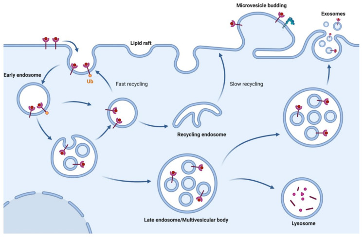
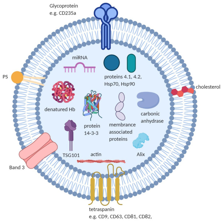
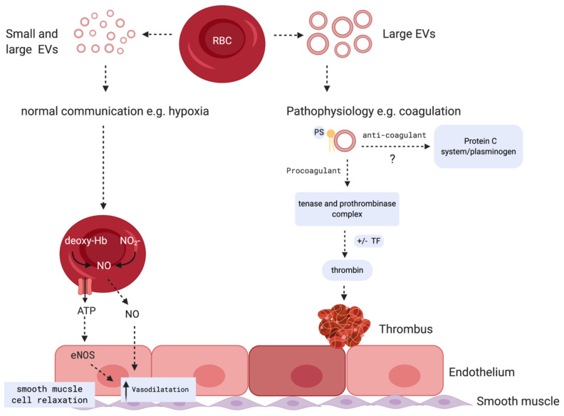
Red blood cells (RBCs) release extracellular vesicles (EVs) including both endosome-derived exosomes and plasma-membrane-derived microvesicles (MVs). RBC-derived EVs (RBCEVs) are secreted during erythropoiesis, physiological cellular aging, disease conditions, and in response to environmental stressors. RBCEVs are enriched in various bioactive molecules that facilitate cell to cell communication and can act as markers of disease. RBCEVs contribute towards physiological adaptive responses to hypoxia as well as pathophysiological progression of diabetes and genetic non-malignant hematologic disease. Moreover, a considerable number of studies focus on the role of EVs from stored RBCs and have evaluated post transfusion consequences associated with their exposure. Interestingly, RBCEVs are important contributors toward coagulopathy in hematological disorders, thus representing a unique evolving area of study that can provide insights into molecular mechanisms that contribute toward dysregulated hemostasis associated with several disease conditions. Relevant work to this point provides a foundation on which to build further studies focused on unraveling the potential roles of RBCEVs in health and disease. In this review, we provide an analysis and summary of RBCEVs biogenesis, composition, and their biological function with a special emphasis on RBCEV pathophysiological contribution to coagulopathy. Further, we consider potential therapeutic applications of RBCEVs.

Keywords: cell-to-cell communication; coagulopathy; exosomes; extracellular vesicles; homeostasis; microparticles; microvesicles; red blood cells.


Conflict of interest statement

The authors declare no conflict of interest.

Figures

Figure 1
Figure 1
Biogenesis of extracellular vesicles (EVs; exosomes and microvesicles): EVs are broadly classified into two categories: exosomes and ectosomes or microvesicles. Exosomes demonstrate a size range between 40 to 150 nm and are generated through a process that involves double invagination of the endosomal membrane to form multivesicular bodies containing intraluminal vesicles. This process is followed by fusion of the multivesicular bodies to the plasma membrane to produce exosomes. By contrast, ectosomes (i.e., microvesicles) and large vesicles are generated by outward budding of the plasma membrane with a size range of 50 to 1000 nm in diameter. (Figure created with BioRender.com).
Figure 2
Figure 2
Mechanisms of RBC-MV biogenesis: MV generation on RBC membrane is predominantly triggered by damaged hemoglobin, protein oxidation, and senescence-associated degradation of band 3-cytoskeletal ankyrin association which result in evagination and vesiculation. Another mechanism involves the alterations in phospholipid distribution in lipid bilayer. Enzymes such as scramblase, calpain, and proteases are activated by oxidative damage or Ca2+ influx via nonspecific cation channels leading to inhibition of flippase and phosphatidylserine externalization, cytoskeletal proteolytic degradation, and band 3 aggregation, resulting in vesiculation. Peroxiredoxin 2 (Prx-2) binding to N-terminal cytoplasmic domain of band 3, phosphorylation (P) and oxidation (O) of band 3 are also indicated [67]. (Figure created with BioRender.com).
Figure 3
Figure 3
Composition of RBCEVs: RBCEVs are reported to contain cytoskeletal proteins (e.g., actin), irreversibly modified Hb, anion transport proteins (e.g., Band 3), glycoproteins (e.g., CD235a), proteins 4.1, 4.2, and 14-3-3, multivesicular body fusion proteins (e.g., Alix, TSG101), membrane-associated proteins (e.g., stomatin (Band 7.2b) and flotillin) and enzymes like carbonic anhydrase. Negatively charged phospholipids (e.g., phosphatidylserine) and other lipid molecules such as cholesterol, and nucleic acid such as miRNA are reported in RBCEVs. (Figure created with BioRender.com).
Figure 4
Figure 4
Potential biological role of RBCEVs: During hypoxia, small and large RBC vesicles carry factors that are responsible for NO production mediated by eNOS, resulting in an increase in vasodilation and smooth muscle cell relaxation (left panel). Large RBC vesicles are reported to play both pro and potential anti-coagulant roles (right panel): MVs mediate procoagulant activities by facilitating assembly of tenase and prothrombinase complexes on phosphatidylserine and promoting thrombin generation. Potential ability of MVs to mediate anticoagulant reactions through their interactions with protein S and activation of anticoagulant protein C system and plasminogen on their surface was reported. This process, in some circumstances, may create an anti-inflammatory and anti-coagulant response based on EV release from certain cells including neutrophils and platelets [116]. (Figure created with BioRender.com).

References

    1. van Niel G., D’Angelo G., Raposo G. Shedding light on the cell biology of extracellular vesicles. Nat. Rev. Mol. Cell Biol. 2018;19:213–228. doi: 10.1038/nrm.2017.125. - DOI - PubMed
    1. Horder E. Blood dust or blood granules: A new constituent of the blood? Lancet. 1899;154:1015. doi: 10.1016/S0140-6736(01)59201-6. - DOI
    1. Nguyen D.B., Ly T.B.T., Bernhardt I. Microvesicles Released from Human Red Blood Cells: Properties and Potential Applications. Volume 10 IntechOpen; Rijeka, Croatia: 2017.
    1. Johnstone R.M., Adam M., Hammond J.R., Orr L., Turbide C. Vesicle formation during reticulocyte maturation. Association of plasma membrane activities with released vesicles (exosomes) J. Biol. Chem. 1987;262:9412–9420. - PubMed
    1. Yáñez-Mó M., Siljander P.R.M., Andreu Z., Zavec A.B., Borràs F.E., Buzas E.I., Buzas K., Casal E., Cappello F., Carvalho J., et al. Biological properties of extracellular vesicles and their physiological functions. J. Extracell Vesicles. 2015;4:27066. doi: 10.3402/jev.v4.27066. - DOI - PMC - PubMed