Review
. 2021 Apr;27(4):367-389.
doi: 10.1261/rna.077271.120. Epub 2020 Dec 29.

A mark of disease: how mRNA modifications shape genetic and acquired pathologies

Affiliations
Review

A mark of disease: how mRNA modifications shape genetic and acquired pathologies

Eliana Destefanis et al. RNA. 2021 Apr.

Abstract

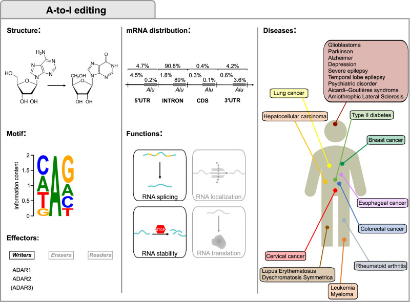
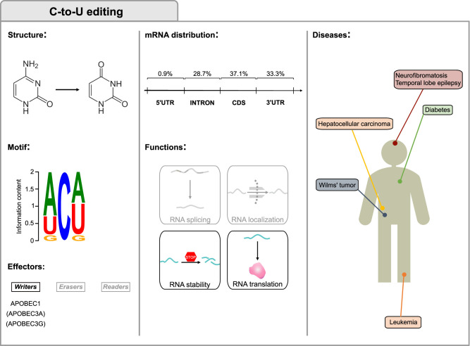
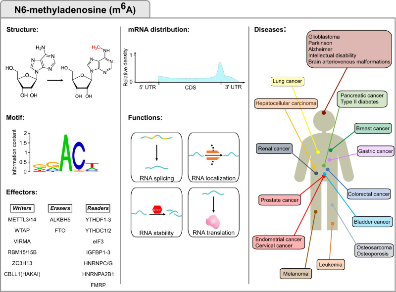
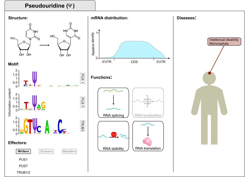
RNA modifications have recently emerged as a widespread and complex facet of gene expression regulation. Counting more than 170 distinct chemical modifications with far-reaching implications for RNA fate, they are collectively referred to as the epitranscriptome. These modifications can occur in all RNA species, including messenger RNAs (mRNAs) and noncoding RNAs (ncRNAs). In mRNAs the deposition, removal, and recognition of chemical marks by writers, erasers and readers influence their structure, localization, stability, and translation. In turn, this modulates key molecular and cellular processes such as RNA metabolism, cell cycle, apoptosis, and others. Unsurprisingly, given their relevance for cellular and organismal functions, alterations of epitranscriptomic marks have been observed in a broad range of human diseases, including cancer, neurological and metabolic disorders. Here, we will review the major types of mRNA modifications and editing processes in conjunction with the enzymes involved in their metabolism and describe their impact on human diseases. We present the current knowledge in an updated catalog. We will also discuss the emerging evidence on the crosstalk of epitranscriptomic marks and what this interplay could imply for the dynamics of mRNA modifications. Understanding how this complex regulatory layer can affect the course of human pathologies will ultimately lead to its exploitation toward novel epitranscriptomic therapeutic strategies.

Keywords: RNA modifications; cancer; epitranscriptomics; human disease; mRNA; posttranscriptional regulation of gene expression.


Figures

FIGURE 1.
FIGURE 1.
A-to-I editing. The first column displays the structures of adenosine and inosine involved in the deamination, the consensus motif and the A-to-I editing main effectors. The motif was obtained by data in Cohen-Fultheim and Levanon (2021) and plotted with WebLogo (Crooks et al. 2004). The central column shows the percentage of editing at nonrepetitive regions and Alu repeats and the functions in mRNA fate. The third column displays A-to-I editing-associated disorders and the organs to which they are associated.
FIGURE 2.
FIGURE 2.
C-to-U editing. The first column displays the structures of cytidine and uridine involved in the deamination, the consensus motif and the C-to-U editing main effectors. The motif was obtained by data in Rosenberg et al. (2011) and plotted with WebLogo (Crooks et al. 2004). The central column shows the percentage of editing in the mRNA regions and the functions in mRNA fate. The third column displays C-to-U editing-associated disorders and the organs to which they are associated. Considering that little is known on the significance of RNA editing by APOBEC3A and APOBEC3G, all features in the figure relate to APOBEC1, and APOBEC3A/APOBEC3G are only mentioned in parentheses.
FIGURE 3.
FIGURE 3.
N6-methyladenosine (m6A) modification. The first column displays the m6A structure, consensus motif and m6A machinery factors. The motif was obtained by data in Linder et al. (2015) and plotted with WebLogo (Crooks et al. 2004). The central column highlights the m6A distribution and functions in mRNA fate, while the third column displays m6A-associated disorders and the organs to which they are associated.
FIGURE 4.
FIGURE 4.
Pseudouridine (Ψ) modification. The first column displays the Ψ structure, consensus motifs and lists the main writers of Ψ in mRNAs. The motif was obtained by data in Carlile et al. (2019) and plotted with WebLogo (Crooks et al. 2004). The central column highlights Ψ distribution (Carlile et al. 2019) and functions in mRNA fate. The third column displays the disorders associated with dysregulated pseudouridylation.
FIGURE 5.
FIGURE 5.
Approaches for the detection of RNA modifications. The figure presents the experimental approaches to the detection of RNA modifications, one per column and left to right: (A) Detection of nonrandom mismatch signatures (RT-signature), (B) capture with antibody, (C) pretreatment with chemical reagents, (D) capture with modification-related enzymes, and (E) detection of modification-specific signals using the Oxford Nanopore Technologies (ONT) platform (ONT-signature). Each column includes the approach schema, with rows below it indicating the approach name, its detection principle, output type, and techniques implementing that approach.

References

    1. Adachi H, Yu YT. 2020. Pseudouridine-mediated stop codon readthrough in S. cerevisiae is sequence context–independent. RNA 26: 1247–1256. 10.1261/rna.076042.120 - DOI - PMC - PubMed
    1. Adachi H, DeZoysa MD, Yu Y-T. 2019a. Detection and quantification of pseudouridine in RNA. Methods Mol Biol 1870: 219–235. 10.1007/978-1-4939-8808-2_17 - DOI - PubMed
    1. Adachi H, De Zoysa MD, Yu Y-T. 2019b. Post-transcriptional pseudouridylation in mRNA as well as in some major types of noncoding RNAs. Biochim Biophys Acta Gene Regul Mech 1862: 230–239. 10.1016/j.bbagrm.2018.11.002 - DOI - PMC - PubMed
    1. Aguilo F, Walsh MJ. 2017. The N6-methyladenosine RNA modification in pluripotency and reprogramming. Curr Opin Genet Dev 46: 77–82. 10.1016/j.gde.2017.06.006 - DOI - PMC - PubMed
    1. Aguilo F, Zhang F, Sancho A, Fidalgo M, Di Cecilia S, Vashisht A, Lee D-F, Chen C-H, Rengasamy M, Andino B, et al. 2015. Coordination of m6A mRNA methylation and gene transcription by ZFP217 regulates pluripotency and reprogramming. Cell Stem Cell 17: 689–704. 10.1016/j.stem.2015.09.005 - DOI - PMC - PubMed

Publication types

MeSH terms