. 2020 Dec 29;117(52):33455-33465.
doi: 10.1073/pnas.2018488117. Epub 2020 Dec 21.

IBEX: A versatile multiplex optical imaging approach for deep phenotyping and spatial analysis of cells in complex tissues

Affiliations

IBEX: A versatile multiplex optical imaging approach for deep phenotyping and spatial analysis of cells in complex tissues

Andrea J Radtke et al. Proc Natl Acad Sci U S A. .

Abstract

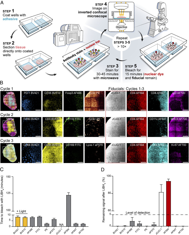
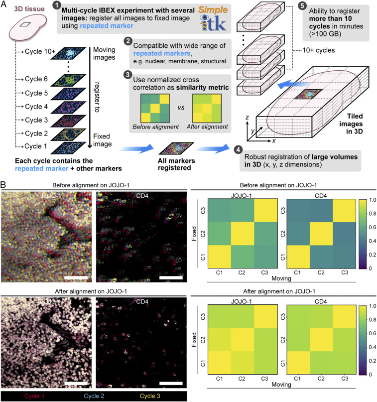
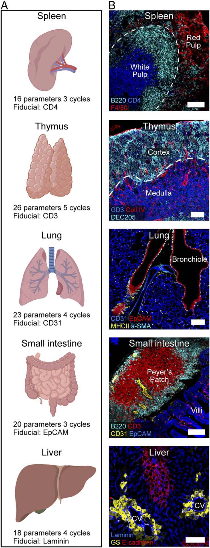
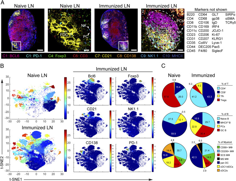
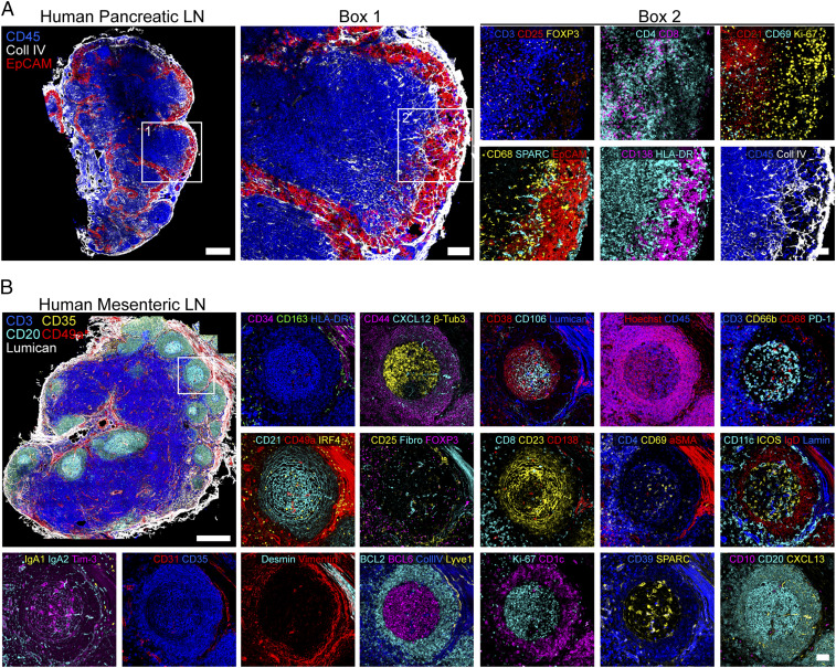
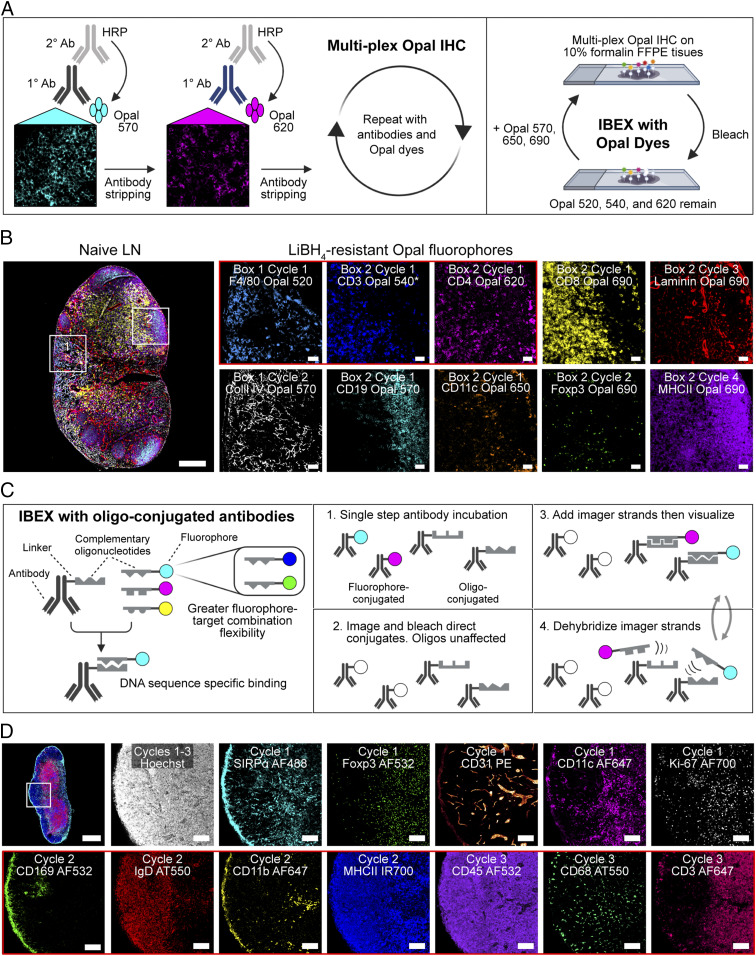
The diverse composition of mammalian tissues poses challenges for understanding the cell-cell interactions required for organ homeostasis and how spatial relationships are perturbed during disease. Existing methods such as single-cell genomics, lacking a spatial context, and traditional immunofluorescence, capturing only two to six molecular features, cannot resolve these issues. Imaging technologies have been developed to address these problems, but each possesses limitations that constrain widespread use. Here we report a method that overcomes major impediments to highly multiplex tissue imaging. "Iterative bleaching extends multiplexity" (IBEX) uses an iterative staining and chemical bleaching method to enable high-resolution imaging of >65 parameters in the same tissue section without physical degradation. IBEX can be employed with various types of conventional microscopes and permits use of both commercially available and user-generated antibodies in an "open" system to allow easy adjustment of staining panels based on ongoing marker discovery efforts. We show how IBEX can also be used with amplified staining methods for imaging strongly fixed tissues with limited epitope retention and with oligonucleotide-based staining, allowing potential cross-referencing between flow cytometry, cellular indexing of transcriptomes and epitopes by sequencing, and IBEX analysis of the same tissue. To facilitate data processing, we provide an open-source platform for automated registration of iterative images. IBEX thus represents a technology that can be rapidly integrated into most current laboratory workflows to achieve high-content imaging to reveal the complex cellular landscape of diverse organs and tissues.

Keywords: high-dimensional imaging; immune system; immunofluorescence; quantitative microscopy; tissue immunity.


Conflict of interest statement

Competing interest statement: J.C. is an employee of BioLegend, which manufactures and sells TotalSeq-A antibodies that are used in one of the workflows described in this paper.

Figures

Fig. 1.
Fig. 1.
IBEX is a high-dimensional, iterative imaging technique. (A) Schematic depicting IBEX protocol. (B) Mice were immunized subcutaneously (s.c.) with 25 µL of SRBCs on day 0 and 7. On day 14, pLN tissue sections were labeled with three separate imaging panels. (Scale bar: 150 µm.) Light refers to bleaching with LiBH4 while the sample was illuminated with a metal halide lamp and DAPI filter. (C) Time required to bleach respective fluorophores using LiBH4. NA, no appreciable loss of signal over multiple hours of LiBH4 exposure. (D) Percentage of fluorophore signal remaining after 15 min of LiBH4 treatment. Data are pooled from two similar experiments and presented as mean ± SEM.
Fig. 2.
Fig. 2.
Image alignment with SimpleITK image registration pipeline. (A) Workflow for SimpleITK image registration pipeline. (B) Confocal images showing JOJO-1 and CD4 from three consecutive IBEX cycles before and after alignment using the nuclear marker JOJO-1 as a fiducial across all three cycles (“C,” Right). CD4 was also repeated and shows cell–cell registration after JOJO-1 alignment. (Scale bar: 50 µm.) Cross-correlation similarity matrices before and after alignment with JOJO-1 for JOJO-1 and CD4 channels. All experiments are representative of at least two similar experiments.
Fig. 3.
Fig. 3.
IBEX in multiple murine organs. (A) IBEX experimental parameters. (B) Confocal images from IBEX experiments in various mouse organs. Liver: central vein (CV) and glutamine synthetase (GS). (Scale bar: 100 µm.) Movies S1–S5 show additional details.
Fig. 4.
Fig. 4.
Visualization and quantification of LN populations using IBEX and histoCAT following immune perturbation. (A) Confocal images of pLNs from naïve and SRBC-immunized mice from 10-cycle (“C” at bottom), 41-parameter IBEX experiments. NK, natural killer; FDC, follicular DC. (Scale bars: left to right, 100 µm, 25 µm, 100 µm, and 50 µm.) (B) t-SNE plots from naïve and immunized LNs identified by Phenograph clustering using segmented cells in histoCAT (naïve, n = 32,091; immune, n = 80,355). Color reflects the cluster ID number (from 1 to 29). Single plots show separation of representative markers into discrete clusters, with color map showing relative expression levels based on Z score-normalized marker intensity values. (C) Phenograph clusters identified by histoCAT were phenotyped based on marker expression and expressed as a proportion of lineage. MΦ, macrophage; SCS, subcapsular sinus; MSM, medullary sinus; dDC, dermal DC. Data are from one experiment and representative of two similar experiments (SI Appendix, Fig. S6 and Movie S6).
Fig. 5.
Fig. 5.
IBEX scales to capture ultra high-content imaging in human tissues. (A) Confocal images from a pancreatic LN with metastatic lesions from a patient with colorectal cancer (4 cycles, 17 parameters). (Scale bars: Left, 500 µm; box 1, 100 µm; box 2, 50 µm). (B) Confocal images from human mesenteric LN (20 cycles, 66 parameters). (Scale bars: 500 µm or 50 µm.) Fibro, fibronectin; Lamin, laminin. Movie S8 shows additional details.
Fig. 6.
Fig. 6.
Incorporation of Opal fluorophores and oligo-conjugated antibodies into IBEX workflow. (A) Opal-plex imaging method. (B) Representative images from a 10-parameter, 4-cycle Opal-plex experiment performed on 5-µm FFPE tissue sections from heavily fixed mouse pLNs. CD3 Opal 540 was present throughout cycles 1 through 4 and served as a fiducial (asterisk). (Scale bars: 50 µm; leftmost panel, 200 µm.) (C) IBEX with oligo-conjugated antibodies method. (D) Confocal images from a 13-parameter, 3-cycle IBEX experiment performed on 20-µm tissue sections from an immunized inguinal mouse LN. Cycle 1: fluorophore-conjugated antibodies. Cycles 2 and 3: oligo-conjugated antibodies, Atto550 (AT550). (Scale bars: 50 µm; Top Left, 400 µm.) Data are representative of three similar experiments (SI Appendix, Fig. S7 and Movie S9).

References

    1. Schubert W., Topological proteomics, toponomics, MELK-technology. Adv. Biochem. Eng. Biotechnol. 83, 189–209 (2003). - PubMed
    1. Schubert W., et al. , Analyzing proteome topology and function by automated multidimensional fluorescence microscopy. Nat. Biotechnol. 24, 1270–1278 (2006). - PubMed
    1. Gerdes M. J., et al. , Highly multiplexed single-cell analysis of formalin-fixed, paraffin-embedded cancer tissue. Proc. Natl. Acad. Sci. U.S.A. 110, 11982–11987 (2013). - PMC - PubMed
    1. Lin J. R., Fallahi-Sichani M., Sorger P. K., Highly multiplexed imaging of single cells using a high-throughput cyclic immunofluorescence method. Nat. Commun. 6, 8390 (2015). - PMC - PubMed
    1. Lin J. R., et al. , Highly multiplexed immunofluorescence imaging of human tissues and tumors using t-CyCIF and conventional optical microscopes. eLife 7, e31657 (2018). - PMC - PubMed

Publication types

Substances

LinkOut - more resources