. 2020 Dec 3:2020:8861557.
doi: 10.1155/2020/8861557. eCollection 2020.

Similar Repair Effects of Human Placenta, Bone Marrow Mesenchymal Stem Cells, and Their Exosomes for Damaged SVOG Ovarian Granulosa Cells

Affiliations

Similar Repair Effects of Human Placenta, Bone Marrow Mesenchymal Stem Cells, and Their Exosomes for Damaged SVOG Ovarian Granulosa Cells

Shuwen Chen et al. Stem Cells Int. .

Abstract

Background: This study is aimed at investigating the repairing effect of mesenchymal stem cells and their exosomes from different sources on ovarian granulosa cells damaged by chemotherapy drugs-phosphoramide mustard (PM).

Methods: In this study, we choose bone marrow mesenchymal stem cells (BMSCs) and human placental mesenchymal stem cells (HPMSCs) for research. Then, they were cocultured with human ovarian granulosa cells (SVOG) injured by phosphoramide mustard (PM), respectively. β-Galactosidase staining, flow cytometry, and Western blot were used to detect the changes in the senescence and apoptosis of SVOG cells before and after their coculture with the above two types of MSCs. Subsequently, exosomes from these two types of MSCs were extracted and added to the culture medium of SVOG cells after PM injury to test whether these two types of exosomes played a role similar to that of MSCs in repairing damaged SVOG cells.

Results: PM treatment-induced apoptotic SVOG cells were significantly decreased after HPMSCs and BMSCs as compared with control group. After coculturing with these two types of MSCs, PM-treated SVOG cells showed significantly reduced senescence and apoptosis proportions as well as cleaved-Caspase 3 expression, and HPMSCs played a slightly stronger role than BMSCs in repairing SVOG cells in terms of the above three indicators. In addition, the ratios of senescent and apoptotic SVOG cells were also significantly reduced by the two types of exosomes, which played a role similar to that of MSCs in repairing cell damages.

Conclusions: The results indicated that BMSCs, HPMSCs, and their exosomes all exerted a certain repair effect on SVOG cells damaged by PM, and consistent repair effect was observed between exosomes and MSCs. The repair effect of exosomes secreted from BMSCs and HPMSCs on the SVOG cells was studied for the first time, and the results fully demonstrated that exosomes are the key carriers for MSCs to play their role.


Conflict of interest statement

The authors declare that they have no competing interests.

Figures

Figure 1
Figure 1
Schematic design of this experiment. (a) The characterization of BMSCs and HPMSCs. The separation and characterization of BMSC-EXO and HPMSC-EXO. (b) Senescence and apoptosis associated with PM-treated SVOG cells. (c) BMSCs or HPMSCs coincubation with PM-treated SVOG cells. (d) BMSCs-EXO or HPMSCs-EXO alone coincubation with PM-treated SVOG cells.
Figure 2
Figure 2
(a) The morphological characteristics of two types of MSCs observed under a microscope (100x): a fibroblast-like long-spindle shape, adherent and vortex-shaped growth, and orderly arrangement. (b) Staining for the induction of adipogenic and osteogenic differentiation of two types of MSCs.
Figure 3
Figure 3
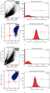
The surface markers of two types of MSCs characterized by flow cytometry: strong expression of CD105 and CD73 and no expression of CD34, CD11b, CD19, CD45, and HLA-DR. (a) BMSC; (b) HPMSC.
Figure 4
Figure 4
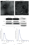
(a) Morphological characteristics of exosomes observed by TEM. (b) Marker protein expression of exosomes secreted from two types of MSCs detected by Western blot. (c) Exosomes characterized by NTA.
Figure 5
Figure 5
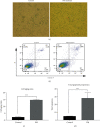
(a, c) The number of dark blue particles in the PM-injured SVOG cells significantly increased (shown by the red arrows), the proportion of senescent cells in the PM treatment group significantly increased, and the difference was statistically significant. ∗∗∗P < 0.001. (b, d) The proportion of apoptotic SVOG cells (the total apoptosis calculated as (Q2 + Q3)) significantly increased after PM injury, and the difference was statistically significant. ∗∗∗P < 0.001.
Figure 6
Figure 6
(a, c) The changes to the senescence of PM-treated SVOG cells after coculturing with BMSCs and HPMSCs (red arrows indicate typical senescent cells). The differences were statistically significant. ∗∗P < 0.01; ∗∗∗P < 0.001. (b, d) The changes to the apoptosis of PM-treated SVOG cells after coculturing with BMSCs and HPMSCs. The calculation formula of the total proportion of apoptosis was (the total apoptosis calculated as (Q2 + Q3)). The differences were statistically significant. P < 0.05; ∗∗∗P < 0.001.
Figure 7
Figure 7
(a) Detection of differences in the protein expression of apoptosis-associated Caspase 3 zymogen and cleaved-Caspase 3 in 4 groups of cells by Western blot. (b) Analysis of difference in gray values. The differences were statistically significant. P < 0.05; ∗∗P < 0.01.
Figure 8
Figure 8
(a, c) The changes to the senescence of PM-treated SVOG cells after the addition of BMSC-EXO and HPMSC-EXO (red arrows indicate typical senescent cells). The differences were statistically significant. ∗∗P < 0.01; ∗∗∗P < 0.001. (b, d) The changes to the apoptosis of PM-treated SVOG cells after the addition of BMSC-EXO and HPMSC-EXO. The calculation formula of the total proportion of apoptosis was (the total apoptosis calculated as (Q2 + Q3)). The differences were statistically significant. P < 0.01; ∗∗∗P < 0.001.
Figure 9
Figure 9
(a) Detection of differences in the protein expression of apoptosis-associated Caspase 3 zymogen and cleaved-Caspase 3 in 4 groups of cells by Western blot. (b) Analysis of difference in gray values. The differences were statistically significant. P < 0.05; ∗∗∗P < 0.001.
Figure 10
Figure 10
(a) Comparison of the effects of BMSCs and their secreted exosomes. (b) Comparison of the effects of HPMSCs and their secreted exosomes.

Similar articles

Cited by

References

    1. Janas P., Kucybała I., Radoń-Pokracka M., Huras H. Telocytes in the female reproductive system: an overview of up-to-date knowledge. Advances in Clinical and Experimental Medicine. 2018;27(4):559–565. doi: 10.17219/acem/68845. - DOI - PubMed
    1. Ozturk S. The translational functions of embryonic poly(A)-binding protein during gametogenesis and early embryo development. Molecular Reproduction and Development. 2019;86(11):1548–1560. doi: 10.1002/mrd.23253. - DOI - PubMed
    1. Tilmann C., Capel B. Mesonephric cell migration induces testis cord formation and Sertoli cell differentiation in the mammalian gonad. Development. 1999;126(13):2883–2890. - PubMed
    1. Hua J., Sidhu K. Recent advances in the derivation of germ cells from the embryonic stem cells. Stem Cells and Development. 2008;17(3):399–412. doi: 10.1089/scd.2007.0225. - DOI - PubMed
    1. Ata B., Seyhan A., Seli E. Diminished ovarian reserve versus ovarian aging: overlaps and differences. Current Opinion in Obstetrics & Gynecology. 2019;31(3):139–147. doi: 10.1097/GCO.0000000000000536. - DOI - PubMed