. 2021 Feb;21(2):134.
doi: 10.3892/etm.2020.9566. Epub 2020 Dec 10.

Poly(ADP-ribose) polymerase-1 inhibitor ameliorates dextran sulfate sodium-induced colitis in mice by regulating the balance of Th17/Treg cells and inhibiting the NF- κ B signaling pathway

Affiliations

Poly(ADP-ribose) polymerase-1 inhibitor ameliorates dextran sulfate sodium-induced colitis in mice by regulating the balance of Th17/Treg cells and inhibiting the NF- κ B signaling pathway

Shuai Peng et al. Exp Ther Med. 2021 Feb.

Abstract

Poly(ADP-ribose) polymerase-1 (PARP-1) plays a critical role in inflammatory pathways. The PARP-1 inhibitor, 5-aminoisoquinolinone (5-AIQ), has been demonstrated to exert significant pharmacological effects. The present study aimed to further examine the potential mechanisms of 5-AIQ in a mouse model of dextran sodium sulfate (DSS)-induced colitis. Colitis conditions were assessed by changes in weight, disease activity index, colon length, histopathology and pro-inflammatory mediators. The colonic expression of PARP/NF-κB and STAT3 pathway components was measured by western blot analysis. Flow cytometry was used to analyze the proportion of T helper 17 cells (Th17) and regulatory T cells (Tregs) in the spleen. Western blot analysis and reverse transcription-quantitative PCR were employed to determine the expression of the transcription factors retinoic acid-related orphan receptor and forkhead box protein P3. The results demonstrated that 5-AIQ reduced tissue damage and the inflammatory response in mice with experimental colitis. Moreover, 5-AIQ increased the proportion of Treg cells and decreased the percentage of Th17 cells in the spleen. Furthermore, following 5-AIQ treatment, the main components of the PARP/NF-κB and STAT3 pathways were downregulated. Collectively, these results demonstrate that the PARP-1 inhibitor, 5-AIQ, may suppress intestinal inflammation and protect the colonic mucosa by modulating Treg/Th17 immune balance and inhibiting PARP-1/NF-κB and STAT3 signaling pathways in mice with experimental colitis.

Keywords: 5-aminoisoquinolinone; T helper 17 cell; poly(ADP-ribose) polymerase-1 inhibitor; regulatory T cell; ulcerative colitis.


Figures

Figure 1
Figure 1
Effects of 5-AIQ on DSS-induced colitis in mice. (A) Body weight, (B) body weight loss, (C) disease activity index score, (D) colon length, (E) histological scores and (F) hematoxylin and eosin staining images (magnification, x200). The results are presented as the mean ± SD (n=10). *P<0.05 vs. control; #P<0.05 vs. DSS group. 5-AIQ, 5-aminoisoquinolinone; DSS, dextran sodium sulfate.
Figure 2
Figure 2
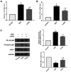
Expression of IL-1β, TNF-α, IκB-α, NF-κB p65 and phospho-NF-κB p65 in colonic tissues. The mRNA levels of (A) IL-1β and (B) TNF-α were measured by reverse transcription-quantitative PCR. (C) The expression levels of IκB-α, NF-κB p65 and phospho-NF-κB p65 were measured by western blot analysis. GAPDH was used as an internal control for grayscale analyses. The results are presented as the mean ± SD (n=10). *P<0.05 vs. control; #P<0.05 vs. DSS group. 5-AIQ, 5-aminoisoquinolinone; DSS, dextran sodium sulfate; phospho, phosphorylated.
Figure 3
Figure 3
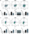
Detection of the frequency of Th17 and Treg cells in the spleen. (A) Flow cytometry and (B) mean averages of the frequency of Th17 cells in the spleen. (C) RT-qPCR analysis of IL-17 A expression. (D) Western blot and (E) RT-qPCR analysis of RORγt expression. (F) Flow cytometry and (G) mean averages of the frequency of Treg cells in the spleen. (H) RT-qPCR analysis of IL-10 expression. (I) Western blot and (J) RT-qPCR analysis of Foxp3 expression. GAPDH was used as an internal control for grayscale analyses. The results are presented as the mean ± SD (n=10). *P<0.05 vs. control; #P<0.05 vs. DSS group. 5-AIQ, 5-aminoisoquinolinone; DSS, dextran sodium sulfate; RT-qPCR. reverse transcription-quantitative PCR; Foxp3, forkhead box protein P3; PE, phycoerythrin; Treg, regulatory T cells; Th17, T helper 17 cells.
Figure 4
Figure 4
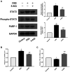
Expression of STAT3, phospho-STAT3, PARP-1, IL-6 and TGF-β1 in colonic tissues. (A) The protein levels of STAT3, phospho-STAT3 and PARP-1 were measured by western blot analysis. GAPDH was used as an internal control for grayscale analyses. (B and C) The mRNA levels of IL-6 and TGF-β1 were measured by RT-qPCR. The results are presented as the means ± SD (n=10). *P<0.05 vs. control; #P<0.05 vs. DSS group.5-AIQ, 5-aminoisoquinolinone; DSS, dextran sodium sulfate.

Similar articles

Cited by

References

    1. Ungaro R, Mehandru S, Allen PB, Peyrin-Biroulet L, Colombel JF. Ulcerative colitis. Lancet. 2017;389:1756–1770. doi: 10.1016/S0140-6736(16)32126-2. - DOI - PMC - PubMed
    1. Kaplan GG, Ng SC. Understanding and preventing the global increase of inflammatory bowel disease. Gastroenterology. 2017;152:313–321.e2. doi: 10.1053/j.gastro.2016.10.020. - DOI - PubMed
    1. de Souza HSP, Fiocchi C, Iliopoulos D. The IBD interactome: An integrated view of aetiology, pathogenesis and therapy. Nat Rev Gastroenterol Hepatol. 2017;14:739–749. doi: 10.1038/nrgastro.2017.110. - DOI - PubMed
    1. Torres J, Colombel JF. Genetics and phenotypes in inflammatory bowel disease. Lancet. 2016;387:98–100. doi: 10.1016/S0140-6736(15)00464-X. - DOI - PubMed
    1. Silva FA, Rodrigues BL, Ayrizono ML, Leal RF. The immunological basis of inflammatory bowel disease. Gastroenterol Res Pract. 2016;2016(2097274) doi: 10.1155/2016/2097274. - DOI - PMC - PubMed