

"Bulb-like" sign: Small bowel closed loop obstruction in incarcerated Spigelian hernia
- PMID: 33376569
- PMCID: PMC7758278
- DOI: 10.1016/j.radcr.2020.12.038
"Bulb-like" sign: Small bowel closed loop obstruction in incarcerated Spigelian hernia
Erratum in
-
Erratum regarding missing declaration of competing interest and patient consent statements in previously published articles.Radiol Case Rep. 2023 Jan 25;18(4):1643-1644. doi: 10.1016/j.radcr.2023.01.017. eCollection 2023 Apr. Radiol Case Rep. 2023. PMID: 36895588 Free PMC article.
Abstract
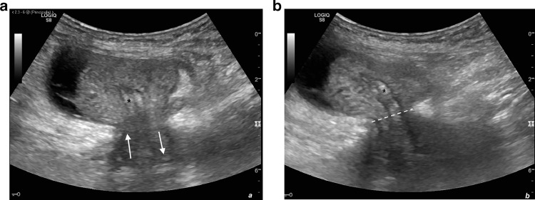
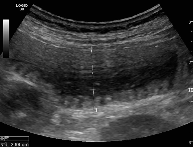
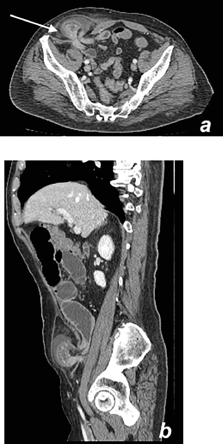
A Spigelian hernia is a rare hernia, making up approximately 0.1% of all abdominal wall hernias. This hernia goes through a defect in the Spigelian fascia which is the part of the transversus abdominis aponeurosis lateral to the rectus muscle, often at the level of the arcuate line, where the fascia is widest and weakest. Clinical diagnosis is difficult in patients without obvious abdominal mass but imaging can be a valuable adjunct in diagnosis. We report the case of a 64-year-old male who presented to our hospital with small bowel obstruction secondary to an incarcerated Spigelian hernia who was pre-operatively diagnosed with ultrasound and computed tomography. At ultrasound and computed tomography a closed loop obstruction in a Spigelian Hernia was detected, resembling on both imaging modalities a "bulb-like" appearance.
Keywords: CT; Incarceration; Small bowel obstruction; Spigelian hernia; Ultrasound.
© 2020 The Authors. Published by Elsevier Inc. on behalf of University of Washington.
Figures
References
Publication types
LinkOut - more resources
Full Text Sources
Other Literature Sources
