. 2020 Nov 16:2020:5097109.
doi: 10.1155/2020/5097109. eCollection 2020.

Synergism and Antagonism of Two Distinct, but Confused, Nrf1 Factors in Integral Regulation of the Nuclear-to-Mitochondrial Respiratory and Antioxidant Transcription Networks

Affiliations

Synergism and Antagonism of Two Distinct, but Confused, Nrf1 Factors in Integral Regulation of the Nuclear-to-Mitochondrial Respiratory and Antioxidant Transcription Networks

Shuwei Zhang et al. Oxid Med Cell Longev. .

Abstract

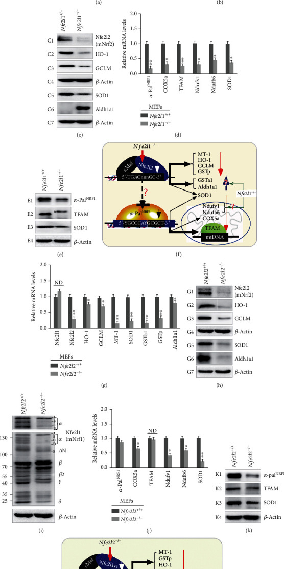
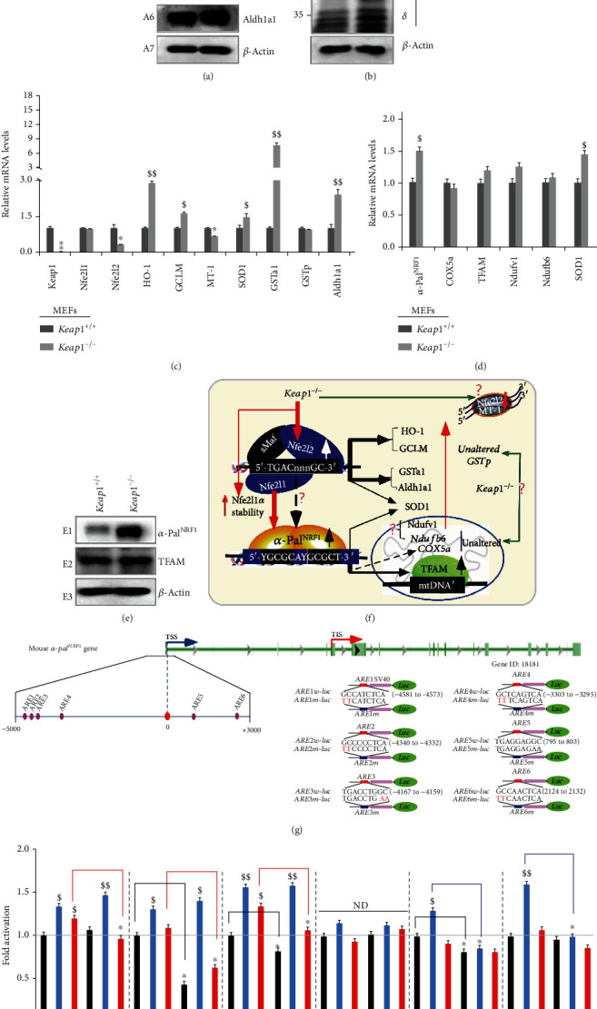
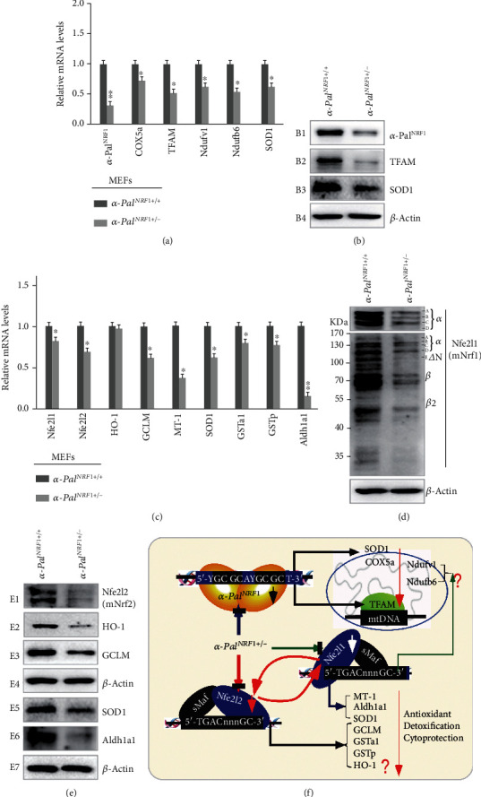
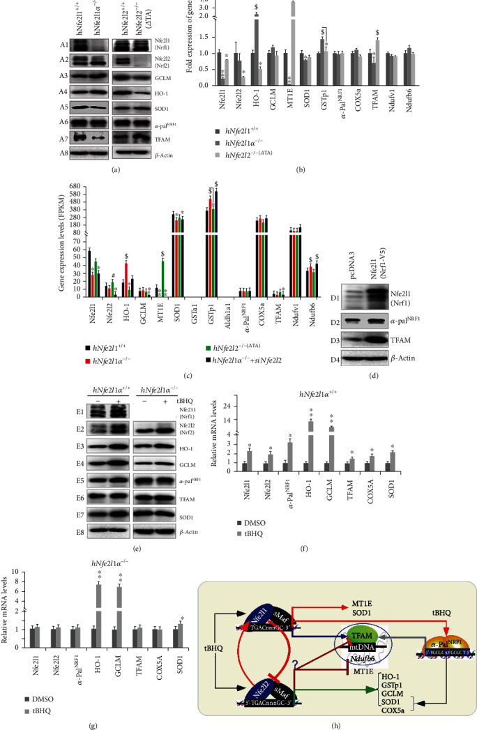
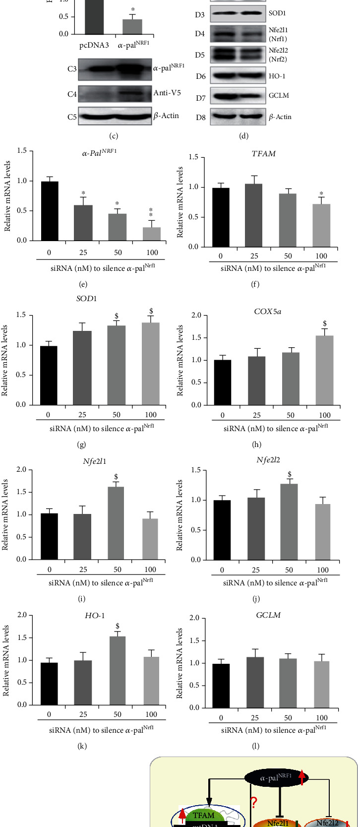
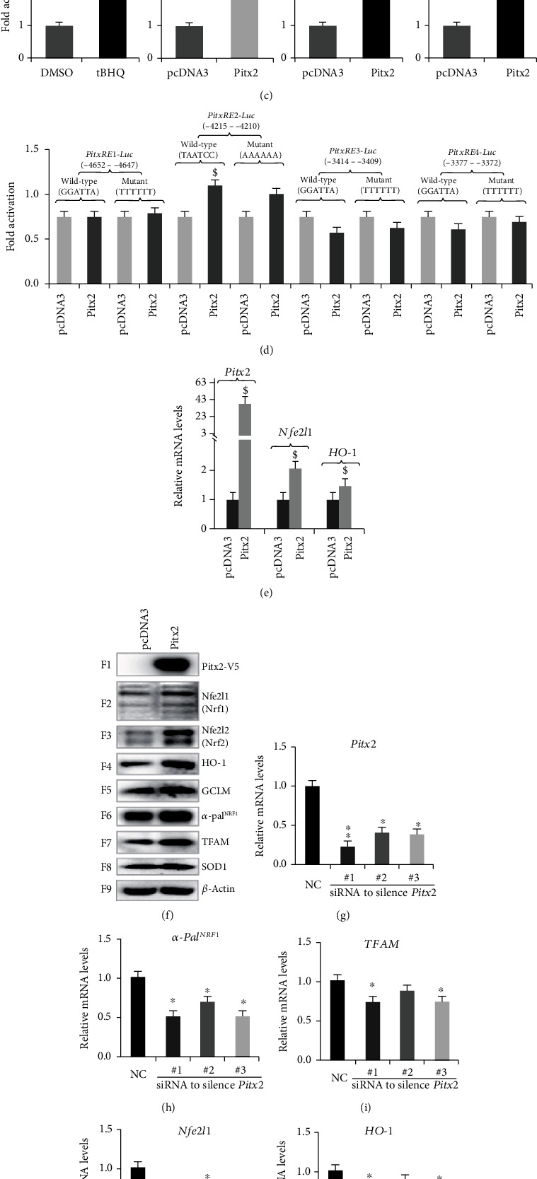
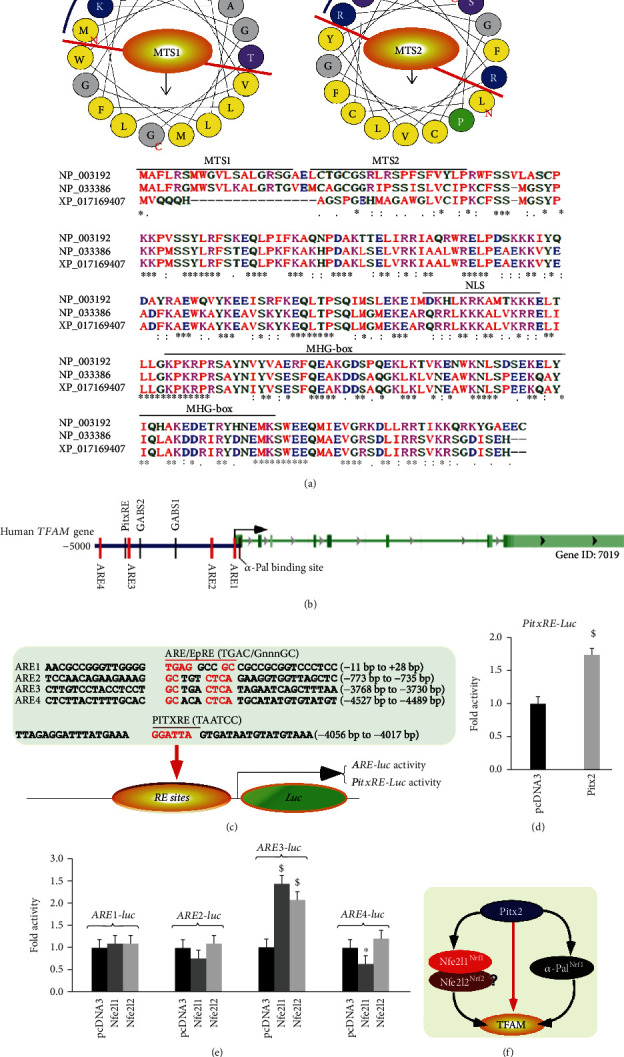
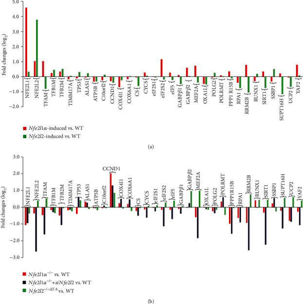
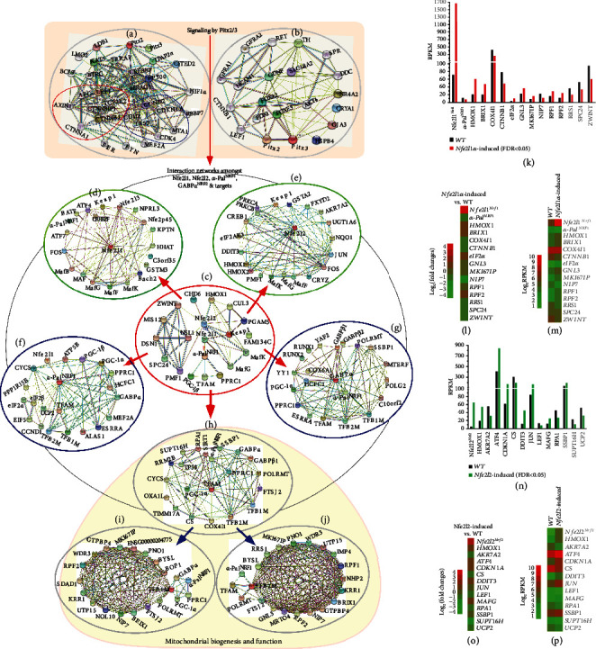
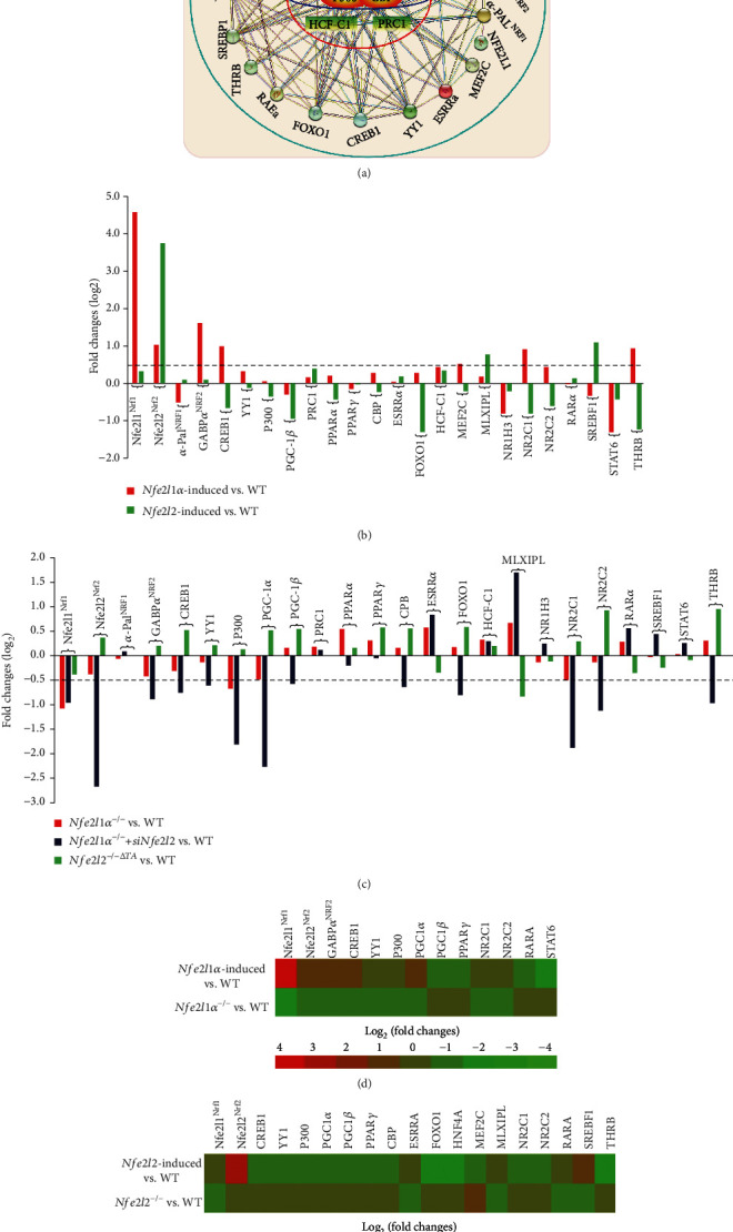
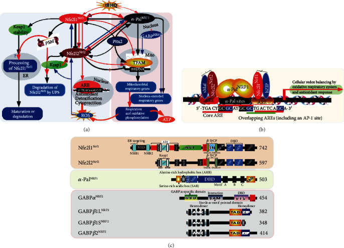
There is hitherto no literature available for explaining two distinct, but confused, Nrf1 transcription factors, because they shared the same abbreviations from nuclear factor erythroid 2-related factor 1 (also called Nfe2l1) and nuclear respiratory factor (originally designated α-Pal). Thus, we have here identified that Nfe2l1Nrf1 and α-PalNRF1 exert synergistic and antagonistic roles in integrative regulation of the nuclear-to-mitochondrial respiratory and antioxidant transcription profiles. In mouse embryonic fibroblasts (MEFs), knockout of Nfe2l1-/- leads to substantial decreases in expression levels of α-PalNRF1 and Nfe2l2, together with TFAM (mitochondrial transcription factor A) and other target genes. Similar inhibitory results were determined in Nfe2l2-/- MEFs but with an exception that both GSTa1 and Aldh1a1 were distinguishably upregulated in Nfe2l1-/- MEFs. Such synergistic contributions of Nfe2l1 and Nfe2l2 to the positive regulation of α-PalNRF1 and TFAM were validated in Keap1-/- MEFs. However, human α-PalNRF1 expression was unaltered by hNfe2l1α-/- , hNfe2l2-/-ΔTA , or even hNfe2l1α-/-+siNrf2, albeit TFAM was activated by Nfe2l1 but inhibited by Nfe2l2; such an antagonism occurred in HepG2 cells. Conversely, almost all of mouse Nfe2l1, Nfe2l2, and cotarget genes were downexpressed in α-PalNRF1+/- MEFs. On the contrary, upregulation of human Nfe2l1, Nfe2l2, and relevant reporter genes took place after silencing of α-PalNRF1, but their downregulation occurred upon ectopic expression of α-PalNRF1. Furtherly, Pitx2 (pituitary homeobox 2) was also identified as a direct upstream regulator of Nfe2l1 and TFAM, besides α-PalNRF1. Overall, these across-talks amongst Nfe2l1, Nfe2l2, and α-PalNRF1, along with Pitx2, are integrated from the endoplasmic reticulum towards the nuclear-to-mitochondrial communication for targeting TFAM, in order to finely tune the robust balance of distinct cellular oxidative respiratory and antioxidant gene transcription networks, albeit they differ between the mouse and the human. In addition, it is of crucial importance to note that, in view of such mutual interregulation of these transcription factors, much cautions should be severely taken for us to interpret those relevant experimental results obtained from knockout of Nfe2l1, Nfe2l2, α-Pal or Pitx2, or their gain-of-functional mutants.


Conflict of interest statement

The authors declare no conflict of interest.

Figures

Figure 1
Figure 1
Distinct effects of Nfe2l1−/−and Nfe2l2−/− on the basal expression of mouse α-PalNRF1, TFAM, and relevant genes in MEFs. (a) The mRNA levels of Nfe2l1 and Nfe2l2, as well as indicated antioxidant genes, were determined by real-time qPCR of Nfe2l1−/− and Nfe2l1+/+ MEFs. The data are shown as mean ± SEM (n = 3 × 3) with significant decreases (p < 0.01, ∗∗p < 0.001) or increases ($, p < 0.01; $$, p < 0.001). (b) Distinct Nfe2l1 isoforms, along with a loading control β-actin, were also examined by Western blotting of Nfe2l1−/− and Nfe2l1+/+ MEFs. (c) Changes in basal protein levels of Nfe2l2 and antioxidant enzymes HO-1, GCLM, SOD1, and Aldh1a1 between Nfe2l1−/− and Nfe2l1+/+ MEFs were also observed. (d) Alterations in the basal mRNA expression levels of α-palNRF1, COX5a, TFAM, Ndufv1, Ndufb6, and SOD1 were unraveled by real-time qPCR of Nfe2l1−/− and Nfe2l1+/+ MEFs. The results are shown as mean ± SEM (n = 3 × 3) with significant decreases (p < 0.01, ∗∗p < 0.001). (e) Altered proteins of α-PalNRF1, TFAM, and SOD1 between Nfe2l1−/− and Nfe2l1+/+ MEFs were visualized by Western blotting. (f) A model is proposed to present possible effects of Nfe2l1−/− on the basal expression of Nfe2l2, α-PalNRF1, TFAM, and relevant genes in MEFs. (g) Basal mRNA levels of Nfe2l1 and Nfe2l2, as well as indicated antioxidant genes, in between Nfe2l2−/− and Nfe2l2+/+ MEFs were also comparatively analyzed. The results are shown as mean ± SEM (n = 3 × 3) with significant decreases (p < 0.01, ∗∗p < 0.001) or ND (no statistical difference). (h) Distinct abundances of Nfe2l2 and antioxidant enzymes in between Nfe2l2−/− and Nfe2l2+/+ MEFs were determined by Western blotting. (i) Altered abundances of distinct Nfe2l1 isoforms in Nfe2l2−/− from Nfe2l2+/+ MEFs were found. (j) The mRNA expression levels of α-PalNRF1, COX5a, TFAM, Ndufv1, Ndufb6, and SOD1 were analyzed by comparing real-time qPCR data from Nfe2l2−/− and Nfe2l2+/+ MEFs. The results are shown as mean ± SEM (n = 3 × 3) with significant decreases (p < 0.01, ∗∗p < 0.001) or ND (no statistical difference). (k) Abundances of α-PalNRF1, TFAM, and SOD1 were compared by immunoblotting of Nfe2l2−/− with Nfe2l2+/+ MEFs. (l) Another model is proposed to present potential influence of Nfe2l2−/− on Nfe2l1, α-PalNRF1, TFAM, and relevant genes (and their proteins) in MEFs.
Figure 2
Figure 2
Distinct roles of key redox control genes in the basal expression of mouse α-PalNRF1, TFAM, and relevant genes. (a) Distinct protein levels of keap1 and Nfe2l2, as well as indicated antioxidant enzymes, in between Keap1−/− and Keap1+/+ MEFs were visualized by Western blotting. (b) Altered abundances of distinct Nfe2l1 isoforms between Keap1−/− and Keap1+/+ MEFs were observed. (c) Basal mRNA levels of keap1, Nfe2l1, and Nfe2l2, as well as indicated antioxidant genes, were determined by real-time qPCR of Keap1−/− and Keap1+/+ MEFs. The data are shown as mean ± SEM (n = 3 × 3) with significant decreases (p < 0.01, ∗∗p < 0.001) or significant increases ($, p < 0.01; $$, p < 0.001). (d) Basal mRNA levels of α-PalNRF1, COX5a, TFAM, Ndufv1, Ndufb6, and SOD1 were also determined as described above. (e) Altered protein levels of α-PalNRF1 and TFAM were revealed by Western blotting of Keap1−/− and Keap1+/+ MEFs. (f) A model is proposed to present effects of Keap1−/− on Nfe2l1, Nfe2l2, α-PalNRF1, TFAM, and related genes (and their proteins) in MEFs. (g) Within the promoter region of mouse α-PalNRF1 gene, the putative ARE sites (each with the core sequence 5′-TGAC/GnnnGC-3′) were marked (as purple dots, left panel). Six distinct ARE-driven reporters (i.e., ARE1 to ARE6-luc) and their respective mutants were constructed into the pGL3-Promoter vector (right panel). (h) Each pair of indicated ARE-luc and mutants was cotransfected with the internal control pRL-TK, together with each of expression constructs for mouse Nfe2l1, Nfe2l2, or empty pcDNA3.1 into RL34 cells for 8 h, before being allowed for 24 h recovery. Subsequently, distinct ARE-driven luciferase activity was measured. The resultant data are shown as mean ± SEM (n = 3 × 3) with significant increases ($, p < 0.01; $$, p < 0.001) or decreases (p < 0.01). ND: no statistical difference.
Figure 3
Figure 3
Distinct effects of mouse α-palNRF1+/- on the expression of Nfe2l1, Nfe2l2, TFAM, and related genes. (a) Altered mRNA levels of α-palNRF1, COX5a, TFAM, Ndufv1, Ndufb6, and SOD1 in α-palNRF1+/- MEFs were compared with their equivalents measured from wild-type (α-palNRF1+/+) cells. The data are shown as mean ± SEM (n = 3 × 3) with significant decreases (p < 0.01, ∗∗p < 0.001). (b) Significant changes in α-PalNRF1, TFAM, and SOD1 proteins were detected by Western blotting of α-palNRF1+/- and α-palNRF1+/+ MEFs. (c) Changes in basal mRNA levels of Nfe2l1 and Nfe2l2, as well as indicated antioxidant genes, were determined by real-time qPCR of α-palNRF1+/- MEFs, when compared with α-palNRF1+/+ MEFs. The results are shown as mean ± SEM (n = 3 × 3) with significant decreases (p < 0.01, ∗∗p < 0.001). (d) Altered abundances of distinct Nfe2l1 isoforms in between α-palNRF1+/- and α-palNRF1+/+ MEFs were also visualized. (e) Altered protein levels of Nfe2l2 and the indicated antioxidant enzymes, such as HO-1, GCLM, SOD1, and Aldh1a1, were further unraveled by Western blotting of α-palNRF1+/- and α-palNRF1+/+ MEFs. (f) A model is proposed to explain distinct effects of α-palNRF1+/- on the nuclear-to-mitochondrial respiratory and antioxidant genes in MEFs.
Figure 4
Figure 4
Distinct changes of human α-PalNRF1 and TFAM in HepG2-derived hNfe2l1α−/− and hNfe2l2−/− cell lines. (a) Distinct protein levels of human Nfe2l1, Nfe2l2, α-PalNRF1, and TFAM as well as other relevant proteins were determined by Western blotting of hNfe2l1α−/−, hNfe2l2−/−, and wild-type HepG2 cells. (b) Basal mRNA expression levels of Nfe2l1, Nfe2l2, α-PalNRF1, TFAM, and other indicated genes were examined by real-time qPCR of hNfe2l1α−/−, hNfe2l2−/−, and wild-type HepG2 cells. The resultant data are shown as mean ± SEM (n = 3 × 3) with significant decreases (p < 0.01, ∗∗p < 0.001) or significant increases ($, p < 0.01; $$, p < 0.001). (c) The FPKM (Reads Per Kilobase per Million mapped reads) value of Nfe2l1, Nfe2l2, α-PalNRF1, TFAM, and other indicated genes were obtained by RNA-sequencing of hNfe2l1α−/−, hNfe2l2−/−, hNfe2l1α−/−+siNfe2l2, and hNfe2l1/2+/+. (d) Different protein levels of Nfe2l1, α-PalNRF1, and TFAM were examined in HepG2 cells that had been transfected with an hNfe2l1 expression construct or empty pcDNA3.1. (e) Distinct inducible alterations in abundances of Nfe2l1, Nfe2l2, α-PalNRF1, TFAM, HO-1, GCLM, and SOD1 were determined by Western blotting of hNfe2l1α+/+ or hNfe2l1α−/− that had been or not been treated with 50μmol/L tBHQ. (f, g) Distinct inducible mRNA levels of Nfe2l1, Nfe2l2, α-PalNRF1, HO-1, GCLM, TFAM, COX5a, and SOD1 were revealed by real-time qPCR of between tBHQ-stimulated lines of hNfe2l1α+/+ cells (f) and hNfe2l1α−/− cells (g). The resultant data are shown as mean ± SEM (n = 3 × 3) with significant increases (p < 0.01, ∗∗p < 0.001). (h) A model is assumed to present cross-talks between human Nfe2l1 and Nfe2l2, along with distinct effects on human α-PalNRF1, TFAM, and other gene expression, particularly upon stimulation by tBHQ.
Figure 5
Figure 5
A negative effect of α-PalNRF1 on Nfe2l1, Nfe2l2, and other antioxidant genes in HepG2 cells. (a) HepG2 cells were cotransfected for 8 h with mNfe2l1-luc reporter and the pRL-TK control, along with an expression construct for mouse α-palNRF1 or an empty pcDNA3.1, and then allowed for a 24 h recovery before the luciferase activity was measured (A1). Total cell lysates were also subjected to identification by Western blotting with distinct antibodies against α-PalNRF1 (A2) or V5 tag (A3). (b) HepG2 cells were cotransfected for 8 h with mNfe2l1-luc (B1) or hNfe2l1-luc (B2), plus pRL-TK, and then treated with 50 μmol/L tBHQ or the DMSO vehicle for 24 h, before being allowed for additional 24 h recovery. Subsequently, these samples were subjected to dual luciferase assays. The results are shown as mean ± SEM (n = 3 × 3) with significant increases ($, p < 0.01). (c) HepG2 cells were cotransfected for 8 h with hNfe2l1-luc (C1) or ARE×6-luc (C2) together with pRL-TK, plus a human α-PalNRF1 expression construct or an empty pcDNA3.1 and then allowed for 24 h recovery from cotransfection, before the reporter activity was measured. The data are shown as mean ± SEM (n = 3 × 3) with significant decreases (p < 0.01). Total cell lysates were also subjected to characterization by Western blotting with distinct antibodies against α-PalNRF1 (C3) or V5 tag (C4). (d) Distinct protein levels of α-PalNRF1, TFAM, SOD1, Nfe2l1, Nfe2l2, HO-1, and GCLM were determined by Western blotting of HepG2 cells that had been transfected with α-PalNRF1 expression plasmid or an empty pcDNA3 vector. (e–l) Distinct changes in mRNA levels of α-PalNRF1 (e), TFAM (f), SOD1 (g), COX5a (h), Nfe2l1 (i), Nfe2l2 (j), HO-1 (k), and GCLM (l) were analyzed by real-time qPCR of HepG2 cells that had been transfected with 0, 25, 50, and 100 nM of siRNA against α-PalNRF1. The resulting data are shown as mean ± SEM (n = 3 × 3) with significant decreases (p < 0.01, ∗∗p < 0.001) or increases ($, p < 0.01). (m) Such siRNA-transfected cell lysates were subjected to Western blotting analysis of distinct protein abundances as indicated. (n) A model is proposed to present disparate effects of α-palNRF1 overexpression or its knockdown on the nuclear-to-mitochondrial respiratory and antioxidant genes in HepG2 cells.
Figure 6
Figure 6
Identification of pitx2 as an upstream regulator of Nfe2l1, besides α-palNRF1. (a) HepG2 cells were cotransfected for 8 h with mNfe2l1-luc (A1) or empty pGL3-Basic (A2), together with the pRL-TK control, plus a Pitx2 expression construct or an empty pcDNA3.1, and then allowed for a 24 h recovery before the luciferase activity was measured. The data are shown as mean ± SEM (n = 3 × 3) with significant increases ($, p < 0.01) or NS (not significance). (b) Putative cis-regulatory binding sites for Pitx2, α-PalNRF1, and AREs within the human Nfe2l1 promoter region were indicated. Various lengths of hNfe2l1-luc were cloned into the pGL3-Basic vector as shown schematically. (c) HepG2 cells were cotransfected for 8 h (i) with hNfe2l1-luc and pRL-TK and also treated for 24 h with 50 μmol/L tBHQ or the DMSO vehicle (C1); (ii) with hNfe2l1-luc (C2), hNfe2l1-luc1 (C3), or hNfe2l1-luc2 (C4), along with the pRL-TK control, plus a Pitx2 expression construct or an empty pcDNA3.1, before being allowed for additional 24 h recovery. The samples were then subjected to dual luciferase assays. The results are shown as mean ± SEM (n = 3 × 3) with significant increases ($, p < 0.01). (d) HepG2 cells were cotransfected for 8 h with each of PitxRE-luc reporters or their mutants, together with pRL-TK plus a Pitx2 expression construct or an empty pcDNA3.1, and then allowed for 24 h recovery. Thereafter, the reporter activity was detected and calculated as mean ± SEM (n = 3 × 3) with significant increases ($, p < 0.01). (e) Distinct mRNA levels of Pitx2, Nfe2l1, and HO-1 were detected by real-time qPCR of HepG2 cells that had been transfected with an expression construct for Pitx2 or an empty pcDNA3.1 vector. (f) Pitx2-expressing HepG2 cells were subjected to Western blotting of Pitx2, Nfe2l1, Nfe2l2, HO-1, GCLM, α-PalNRF1, TFAM, and SOD1. (g–n) Distinct mRNA levels of Pitx2 (g), α-PalNRF1 (h), TFAM (i), Nfe2l1 (j), HO-1 (k), GCLM (l), COX5a (m), and SOD1 (n) were determined by real-time qPCR analysis of HepG2 cells that had been transfected with three different siRNAs against Pitx2. The results are shown as mean ± SEM (n = 3 × 3) with significant decreases (p < 0.01, ∗∗p < 0.001). (o) A model is proposed for effects of Pitx2 on Nfe2l1, Nfe2l2, α-PalNRF, TFAM, and other genes in HepG2 cells.
Figure 7
Figure 7
Transcriptional regulation of TFAM by Nfe2l1, Nfe2l2, and Pitx2. (a) Two similar α-helical structural wheels were formed by successive mitochondria-targeting sequences MTS1 (aa 1-18) and MTS2 (aa 21-38). Basic arginine and lysine residues were placed on blue backgrounds; nucleophilic serine and threonine residues are on purple backgrounds; an unamiable proline residue was on a green background, and all other hydrophobic amino acids were on yellow backgrounds, except for small alanine and glycine on grey backgrounds. The lower panel shows an alignment of three amino-acid sequences of human TFAM (NP_003192) and mouse TFAMs (NP_033386 and XP_017169407), in which, MTS1, MTS2, nuclear localization signal (NLS), and DNA-binding MHG-box were indicated. (b) The putative consensus ARE sites and other cis-regulatory binding sites for Pitx2, α-PalNRF1, or GABP within the human TFAM gene promoter region were indicated. (c) Four distinct ARE-driven (i.e., ARE1-luc to ARE4-luc) and another Pitx2RE-luc reporters were constructed into the pGL3-Promoter vector. (d) HepG2 cells were cotransfected with PitxRE-luc and pRL-TK, plus a Pitx2 expression plasmid or an empty pcDNA3.1, and then allowed for 24 h recovery before the luciferase activity was measured. The data are shown as mean ± SEM (n = 3 × 3) with significant increase ($, p < 0.01). (e) HepG2 cells were cotransfected with each of ARE1-luc to ARE4-luc, together with pRL-TK plus an expression construct for Nfe2l1, Nfe2l2, or an empty pcDNA3.1, and then allowed for 24 h recovery, before such ARE-driven activity was detected. The resultant data are shown as mean ± SEM (n = 3 × 3) with significant increases ($, p < 0.01) or decreases (p < 0.01). (f) A model is proposed to explain the transcriptional regulation of TFAM by Nfe2l1, Nfe2l2, Pitx2, and α-PalNRF1 in HepG2 cells.
Figure 8
Figure 8
Distinct or even opposite contributions of Nfe2l1 and Nfe2l2 to expression of TFAM, TFB1M, TFB2M, and other critical genes for the nuclear-to-mitochondrial communication. (a) 33 of differential expression genes (DEGs) were selected from RNA-sequencing of either Nfe2l1α-induced and Nfe2l2-induced cell lines versus their WT controls (by the value of FDR < 0.05). (b) Those gene expression levels were also determined by RNA-sequencing of Nfe2l1α−/−, Nfe2l1α−/−+siNfe2l2, and Nfe2l2-/-ΔTA cell lines. Their values were then calculated by Log2 (fold change), relative to their equivalents of wild-type (WT) control cells.
Figure 9
Figure 9
A hierarchical interaction network integrated with those relevant genes involved in the nuclear-to-mitochondrial communication. (a, b) Two interaction subnetworks with Pitx2- and Pitx3-regulated genes. (c–e) A core subnetwork of redox-relevant genes regulated by Nfe2l1 and/or Nfe2l2, together with Nfe2l1- and/or Nfe2l2-interactors in additional two extended subnetworks. (f–j) Five subnetworks are monitored by α-PalNRF1, GABPαNRF2, TFAM, TFB1M, and/or TFB2M, which are key players as the nuclear controls of mitochondrial biogenesis and function. (k–m) Differentially expressed genes (DEGs) are contributed by the Nfe2l1α-inducible expression. Their FPKM values (FDR < 0.05) were obtained from RNA-sequencing of Nfe2l1α-induced cells versus WT cells. Such DEGs were also shown by two distinct heat maps, as scaled in different ways. (n–p) 14 of DEGs are regulated by Nfe2l2-inducible expression, which are also shown in two distinct fashions as described above.
Figure 10
Figure 10
Nfe2l1 and Nfe2l2 contribute to differential expression of PGC-1α/β and associated genes. (a) The PGC-1α/β-centered interaction network with distinct families of 21 transcription factors and additional 4 cofactors, which was constructed by the STRING tool (https://string-db.org/). (b) The Log2 (fold change) values of those indicated genes were calculated by comparison of RNA-sequencing data obtained from either Nfe2l1α-induced or Nfe2l2-induced cells with equivalent wild-type (WT) controls. (c) Similar Log2 (fold change) values of the above-described genes were also calculated by comparison of RNA-sequencing data obtained from Nfe2l1α−/−, Nfe2l1α−/−+siNfe2l2, or Nfe2l2-/-ΔTA cell lines versus their equivalent WT controls (FDR < 0.05). (d) A heat map shows differential expression levels of Nfe2l1α-affected genes determined by comparison of either Nfe2l1α−/− or Nfe2l1α-induced cells with the respective WT controls (FDR < 0.05). Such different colored genes were scaled by their Log2 values. (e) Another heat map of 19 Nfe2l2-affected genes, which were determined by comparison of Nfe2l2-/-ΔTA or Nfe2l2-induced cells with their respective WT controls (FDR < 0.05).
Figure 11
Figure 11
An integral model to interpret coordinated regulation of distinct cellular respiratory and antioxidant gene transcription networks. (a) A model is based on our experimental evidence, to give a better understanding of regulatory cross-talk among Nfe2l1Nrf1, Nfe2l2Nrf2, α-PalNRF1, and Pitx2, all targeting TFAM. During the nuclear-to-mitochondrial communication, Nfe2l1Nrf1 makes opposing contributions to bidirectional regulation of Nfe2l2Nrf2 and α-PalNRF1 by itself and target proteasome (PSM) at two distinct layers. Nfe2l2Nrf2 can determine posttranscriptional regulation of Nfe2l1Nrf1 and α-PalNRF1, but the detailed mechanism remains unclear. In turn, mouse α-PalNRF1 contributes to positive regulation of Nfe2l1Nrf1 and Nfe2l2Nrf2, although no canonic GC-rich α-Pal-binding sites exist in these mouse CNC-bZIP gene promoter regions (Table S2). Contrarily, human α-PalNRF1 makes a negative contribution to transcriptional expression of Nfe2l1Nrf1 and Nfe2l2Nrf2 (the latter Nfe2l2Nrf2 is dominantly negatively regulated by human Nfe2l1Nrf1), albeit all three factors are activated by redox inducer tBHQ. In addition to negative regulation of Nfe2l2Nrf2 by Keap1, the adaptor subunit of Cullin 3-based E3 ubiquitin ligase can also make a positive contribution to transcriptional regulation of mouse Nfe2l2Nrf2, rather than Nfe2l1Nrf1, as found in MEFs. However, human Nfe2l1Nrf1 is essential for stabilization of Keap1, but it is unknown whether this adaptor protein is involved in the proteolytic processing of Nfe2l1Nrf1. Notably, the nucleus-controlled mitochondrial respiratory and oxidative phosphorylation are also a primary source of ROS in cells, which triggers activation of Nfe2l1Nrf1, Nfe2l2Nrf2, and α-PalNRF1 to certain extents so that cellular redox homeostasis is maintained at a steady state. Besides, GABPNRF2 is also required for this process, but possible cross-talks of this ETS family factor with Nfe2l1Nrf1 and Nfe2l2Nrf2 are not yet identified here. The “M”-marked arrowheads indicate those activity in the mouse but not in the human; such distinction was also shown (in Figure S2). (b) Schematic explanation of the intracellular redox homeostasis balanced by an oxidative respiratory system and another antioxidant cytoprotective response. Most of ARE-driven genes are transcriptionally regulated by distinct functional heterodimers of either Nfe2l1Nrf1or Nfe2l2Nrf2 with sMaf or other bZIP proteins, whilst most of the nucleus-encoded mitochondrial respiratory genes are controlled predominantly by α-PalNRF1 homodimers. Of note, the GC-enriched α-Pal-binding site is overlapped with the -GC-motif of ARE-core sequences (each of which contains an AP-1 site). This implies synergistic and/or antagonistic regulatory effects of Nfe2l1Nrf1, Nfe2l2Nrf2, and α-PalNRF1 on certain expression of distinct cognate target genes. (c) Schematic representation of distinct structural domains of Nfe2l1Nrf1, Nfe2l2Nrf2, and α-PalNRF1, as well as GABPαNRF2, GABPβ1LNRF2, GABPβ1SNRF2, and GABPβ2NRF2. Of note, all domains and motifs of Nfe2l1Nrf1 and Nfe2l2Nrf2 were defined (37), but neither have no homology with α-PalNRF1, GABPαNRF2, and GABPβNRF2. Distinct domains and motifs of these nuclear respiratory factors are identified by bioinformatic analysis of their amino-acid sequences. ANK: ankyrin repeats; DBD: DNA-binding domain; ER: endoplasmic reticulum; ETS: E26 transformation specific; GSD: GABPα-specific domain; NLS: nuclear localization signal; Mito: mitochondria; SAM: sterile α-motif pointed domain; TAD: transactivation domain.

Similar articles

Cited by

References

    1. Gould S. B., Garg S. G., Martin W. F. Bacterial vesicle secretion and the evolutionary origin of the eukaryotic endomembrane system. Trends in Microbiology. 2016;24(7):525–534. doi: 10.1016/j.tim.2016.03.005. - DOI - PubMed
    1. Griffiths G. Cell evolution and the problem of membrane topology. Nature Reviews. Molecular Cell Biology. 2007;8(12):1018–1024. doi: 10.1038/nrm2287. - DOI - PubMed
    1. Amigo I., da Cunha F. M., Forni M. F., et al. Mitochondrial form, function and signalling in aging. The Biochemical Journal. 2016;473(20):3421–3449. doi: 10.1042/BCJ20160451. - DOI - PubMed
    1. Murray A. J., Horscroft J. A. Mitochondrial function at extreme high altitude. The Journal of Physiology. 2016;594(5):1137–1149. doi: 10.1113/JP270079. - DOI - PMC - PubMed
    1. Gray M. W. Mitochondrial Evolution. Cold Spring Harbor Perspectives in Biology. 2012;4(9):p. a011403. doi: 10.1101/cshperspect.a011403. - DOI - PMC - PubMed