

Gallbladder-duodenal fistula detected by ultrasound - a case report
- PMID: 33376600
- PMCID: PMC7768895
- DOI: 10.15557/JoU.2020.0036
Gallbladder-duodenal fistula detected by ultrasound - a case report
Abstract
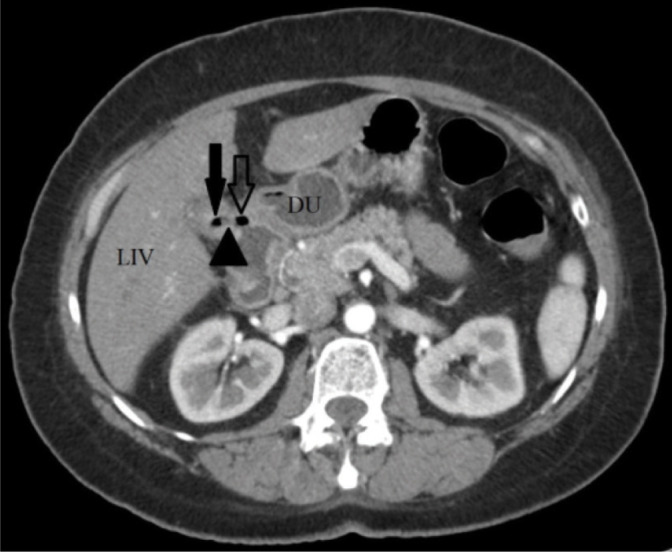
Gallbladder-duodenal (cholecystoduodenal) fistula is an uncommon bilioenteric fistula between the gallbladder and the duodenum. It usually occurs following a chronic case of cholecystitis upon which the gallbladder adheres to the adjacent duodenum, and a stone penetrates through the wall. The case presented herein is that of a gallbladder-duodenal fistula detected primarily with the use of ultrasound imaging, and subsequently confirmed by computed tomography. The patient is a 54-year-old woman who was admitted with upper abdominal pain. The fistula was caused by chronic cholecystitis, however no gallstones were present in the duodenum. Surgical management was undertaken for the patient, and the recovery was uneventful.
Keywords: cholecystitis; chronic; gallbladder-duodenal fistula; gallstones.
© Polish Ultrasound Society.
Figures


References
-
- Feferman Y, Bard V, Aviran N, Stein M, Kashtan H, Sadot E: An unusual presentation of cholecystoduodenal fistula: massive upper gastrointestinal bleeding. J Gastrointest Dig Syst 2015; 5: 314.
-
- Glenn F, Reed C, Grafe WR: Biliary enteric fistula. Surg Gynecol Obstet 1981; 153: 527–531. - PubMed
-
- Shaffer EA: Gallstone disease: epidemiology of gallbladder stone disease. Best Pract Res Clin Gastroenterol 2006; 20: 981–996. - PubMed
LinkOut - more resources
Full Text Sources