

SOX1 and PAX1 Are Hypermethylated in Cervical Adenocarcinoma and Associated with Better Prognosis
- PMID: 33376722
- PMCID: PMC7738792
- DOI: 10.1155/2020/3981529
SOX1 and PAX1 Are Hypermethylated in Cervical Adenocarcinoma and Associated with Better Prognosis
Abstract
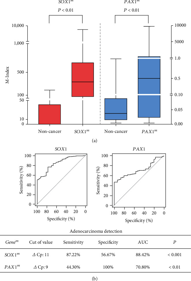
Background: The increased risk and poor survival outcome of cervical adenocarcinoma (CAC) demand for effective early diagnostic biomarkers that can predict the disease progression and outcome. The purpose of this study was to investigate the value of methylation status of SOX1 and PAX1 in the detection and prognosis of CAC.
Methods: We performed a quantitative methylation-specific polymerase chain reaction in 205 cervical paraffin-embedded specimens (175 CACs, 30 noncancer cervical tissues). Overall and progression-free survival (OS and PFS, respectively) rates were calculated and compared using the Kaplan-Meier method. The prognostic value of SOX1m and PAX1m on CAC patients was assessed by the Cox regression model. A mathematical formula combining SOX1m , PAX1m , and age was constructed for survival prediction.
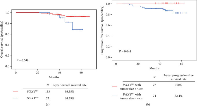
Results: The methylation status of SOX1 and PAX1 was higher in CAC tissues than in noncancer cervical tissues. In addition, SOX1m -positive CAC patients showed a higher 5-year OS rate than SOX1m -negative patients. In CAC patients with smaller tumor size (<4 cm), the PAX1m -positive group showed a higher 5-year PFS rate than the PAX1m -negative group. In the algorithm combining SOX1m , PAX1m , and age, the low-risk group showed a better 5-year OS and PFS rate than the high-risk group.
Conclusion: SOX1 and PAX1 methylation levels are higher in CAC than in normal cervical tissues and are potential biomarkers for monitoring CAC prognosis.
Copyright © 2020 Zitong Zhao et al.
Conflict of interest statement
The authors declare that they have no competing interests.
Figures

References
-
- Jemal A., Bray F., Center M. M., Ferlay J., Ward E., Forman D. Global cancer statistics. CA: a cancer journal for clinicians. 2015;65(2):87–108. - PubMed
-
- Drescher C. W., Hopkins M. P., Roberts J. A. Comparison of the pattern of metastatic spread of squamous cell cancer and adenocarcinoma of the uterine cervix. Gynecologic oncology. 1989;33(3):340–343. - PubMed
-
- Eifel P. J., Morris M., Oswald M. J., Taylor Wharton J., Delclos L. Adenocarcinoma of the uterine cervix. Prognosis and patterns of failure in 367 cases. Cancer. 1990;65(11):2507–2514. - PubMed
MeSH terms
Substances
LinkOut - more resources
Full Text Sources
Medical
Molecular Biology Databases
