. 2020 Dec 21:8:tkaa036.
doi: 10.1093/burnst/tkaa036. eCollection 2020.

Co-Cr-Mo-Cu alloys for clinical implants with osteogenic effect by increasing bone induction, formation and development in a rabbit model

Affiliations

Co-Cr-Mo-Cu alloys for clinical implants with osteogenic effect by increasing bone induction, formation and development in a rabbit model

Jingzhu Duan et al. Burns Trauma. .

Abstract

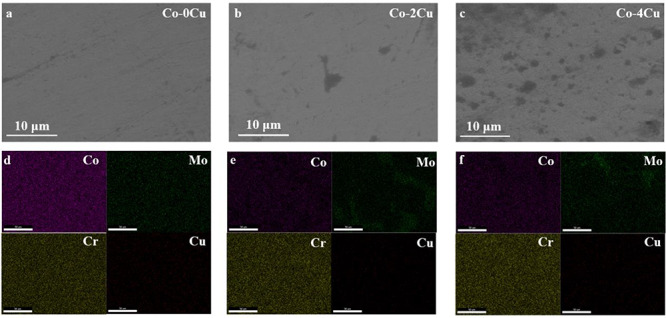
Background: Co-Cr-Mo alloy has been widely used in clinical implants because of its excellent mechanical and anti-corrosion properties, but there is an urgent need to address its disadvantages, such as implant-related infections and implant loosening. We synthesized Co-Cr-Mo-Cu (Co-Cu) alloys with different Cu contents to modify implant performance to be suitable as a bone-compatible implant material.

Methods: Microstructure, phase content and mechanical properties of the Co-Cr-Mo alloy were characterized. Histological and immunohistochemical analyses were performed after implantation in rabbits. The experimental alloy was implanted on the lateral side of the lower tibial condyle and the tibial nodule.

Results: Phase content and mechanical properties revealed that the crystallographic structure and wear resistance were changed. Experimental implantation results demonstrated that osteogenic capability was markedly enhanced, ascribed to the excellent antibacterial and osseointegration capacities of Cu phases, and with the release of Cu ions. In particular, Co-Cu alloy containing 2 wt% Cu exhibited the best osteogenic performance among all samples.

Conclusions: The results indicated that osteogenic performance of the Co-Cr-Mo alloy could be enhanced by adding Cu. In particular, the Co-2Cu alloy exhibited the best properties according to both immunohistochemical and histological analyses. Our study not only provides deep insight into the osteogenic effect of Cu but presents a new Co-Cu alloy for clinical implants.

Keywords: Clinical application; Co-Cr-Mu-Cu alloy; Cu ion; Implantable operation; Osteogenic capability.


Figures

Figure 1.
Figure 1.
Phase characterization of three different Co-Cu alloys (by X-ray diffraction), where it can be seen that the hexagonal closepacked structure (HCP) and face center cubic (FCC) phases and the FCC γ-Co phase could modify ductility and deformability of Co-Cu alloys
Figure 2.
Figure 2.
Microstructure of Co-Cu alloys with different Cu content. The second phase structure showed a clear difference. (a–c) High resolution-scanning electron microscope images of different Co-Cu alloys. (d–f) The corresponding energy-scattering spectrum mapping images, scale bar: 50 μm
Figure 3.
Figure 3.
Element characterizations (X-ray photoelectron spectroscopy) of three different Co-Cu alloys. The C1s and O1s peaks observed were ascribed to the surface segregation behavior of the Co-Cu alloys
Figure 4.
Figure 4.
Tensile stress–strain curves of the different Co-Cu alloys. The Co-Cu alloys showed improved ductility
Figure 5.
Figure 5.
The friction coefficient and abrasion loss of the different Co-Cu alloys. The enhancement of abrasion loss performance was ascribed to the large carbide phases and inhomogeneous element distribution (*p < 0.05)
Figure 6.
Figure 6.
Ion release of Co-Cu alloys in 0.9% NaCl solution after 1-day immersion. Co, Cr and Cu were detected with all Co-Cu alloys. The ion concentrations were much lower than reported values, which makes it less likely that the released ions would result in cell toxicity
Figure 8.
Figure 8.
Immunohistochemical analysis of Co-Cu alloy implantation (bone morphogenetic protein-2), which might reflect the mesenchymal cells differentiating into cartilage cells and thereby forming new bone, with a composition including osteoblasts, osteoclasts and bone marrow tissue (scale bar: 20 μm) (a) Ti alloy; (b) Co-0Cu alloy; (c) Co-2Cu alloy; (d) Co-4Cu alloy
Figure 9.
Figure 9.
Immunohistochemical analysis of Co-Cu alloy implantation (insulin-like growth factor), which could reflect the synthesis of DNA and RNA in osteoblasts, participating in bone remodeling and bone metabolism for bone resorption (scale bar: 20 μm). (a) Ti alloy; (b) Co-0Cu alloy; (c) Co-2Cu alloy; (d) Co-4Cu alloy
Figure 10.
Figure 10.
The average optical densities for: (a) bone morphogenetic protein-2 (BMP-2) for Ti and Co-Cu alloys in the 1-, 2-, 3- and 4-week groups; and (b) insulin-like growth factor (IGF-1) for Ti and Co-Cu alloys in the 1-, 2-, 3- and 4-week groups. IGF-1 and BMP-2 expression measured by enzyme-linked immunosorbent assay: (c) BMP-2 for Ti and Co-Cu alloys in 1-, 2-, 3- and 4-week groups; and (d) IGF-1 for Ti and Co-Cu alloys in 1-, 2-, 3- and 4-week groups (*p < 0.05; ** p < 0.01)
Figure 11.
Figure 11.
Histological structures with Co-Cu alloys. After Masson’s trichrome staining, bone trabeculae exhibited a blue-stained structure of varying depth. Bone cells appear as irregular white dots, fibrous tissue is pink-stained and new bone is not stained (scale bar: 100 µm). (a) Ti alloy; (b) Co-0Cu alloy; (c) Co-2Cu alloy; (d) Co-4Cu alloy
Figure 12.
Figure 12.
Statistical results of bone–implant contact (BIC) and mineral apposition rate (MAR) characterization for the different alloys. (a) BIC measurements for Ti and Co-Cu alloys in the 4-, 8- and 12-week groups. (b) MAR measurements for Ti and Co-Cu alloys in the 4-, 8- and 12-week groups (*p < 0.05; ** p < 0.01)
Figure 13.
Figure 13.
Mineral apposition rate characterization of the different Co-Cu alloys. Using fluorescent labeling of the position of bone deposition at different time points and different directions, the bone formation distance in this study period was measured (scale bar: 500 µm). (a) Ti alloy; (b) Co-0Cu alloy; (c) Co-2Cu alloy; (d) Co-4Cu alloy
Figure 7.
Figure 7.
The osseous connection between the implant alloy and bone tissue. (a) Scheme of the corresponding animal experiment. (b) Radiologic examination for implant alloy and bone tissue at 4, 8 and 12 weeks. It can be seen that, in many cases, the lamellar and cortical bone is filled with new bone tissue and the density of this is close to that of mature bone

Similar articles

Cited by

References

    1. Hodgson AWE, Kurz S, Virtanen S, Fervel V, Olsson COA, Mischler S. Passive and transpassive behaviour of CoCrMo in simulated biological solutions. Electrochim Acta. 2014;49:2167–78.
    1. Xiang DD, Wang P, Tan XP, Chandra S, Wang C, Nai MLS, et al. Anisotropic microstructure and mechanical properties of additively manufactured Co–Cr–Mo alloy using selective electron beam melting for orthopedic implants. Mater Sci Eng A Struct Mater. 2019;765:138270 10.1016/j.msea.2019.138270. - DOI
    1. Igual Muñoz A, Casabán Julián L. Influence of electrochemical potential on the tibocorrosion behaviour of high carbon CoCrMo biomedical alloy in simulated body fluids by electrochemical impedance spectroscopy. Electrochim Acta. 2010;55:5428–39.
    1. Wei D, Koizumi Y, Takashima T, Nagasako M, Chiba A. Fatigue improvement of electron beam melting-fabricated biomedical co–Cr–Mo alloy by accessible heat treatment. Mater Res Lett. 2018;6:93–9.
    1. Shah F, Omar O, Suska F, Snis A, Matic A, Emanuelsson L, et al. Long-term osseointegration of 3D printed CoCr constructs with an interconnected open-pore architecture prepared by electron beam melting. Acta Biomater. 2016;36:296–309. - PubMed