. 2020 Oct 13;2(2):fcaa150.
doi: 10.1093/braincomms/fcaa150. eCollection 2020.

The adult human subventricular zone: partial ependymal coverage and proliferative capacity of cerebrospinal fluid

Affiliations

The adult human subventricular zone: partial ependymal coverage and proliferative capacity of cerebrospinal fluid

Sophia F A M de Sonnaville et al. Brain Commun. .

Abstract

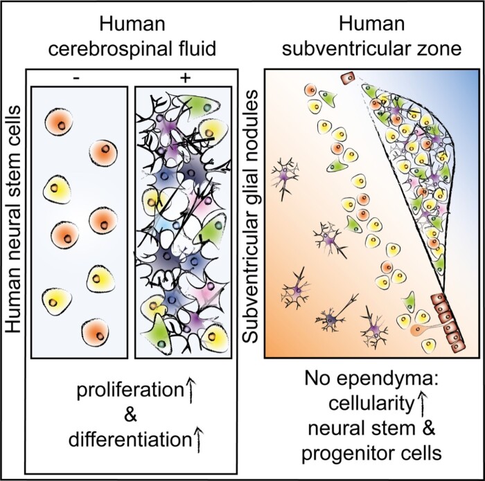
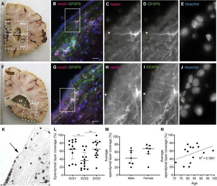
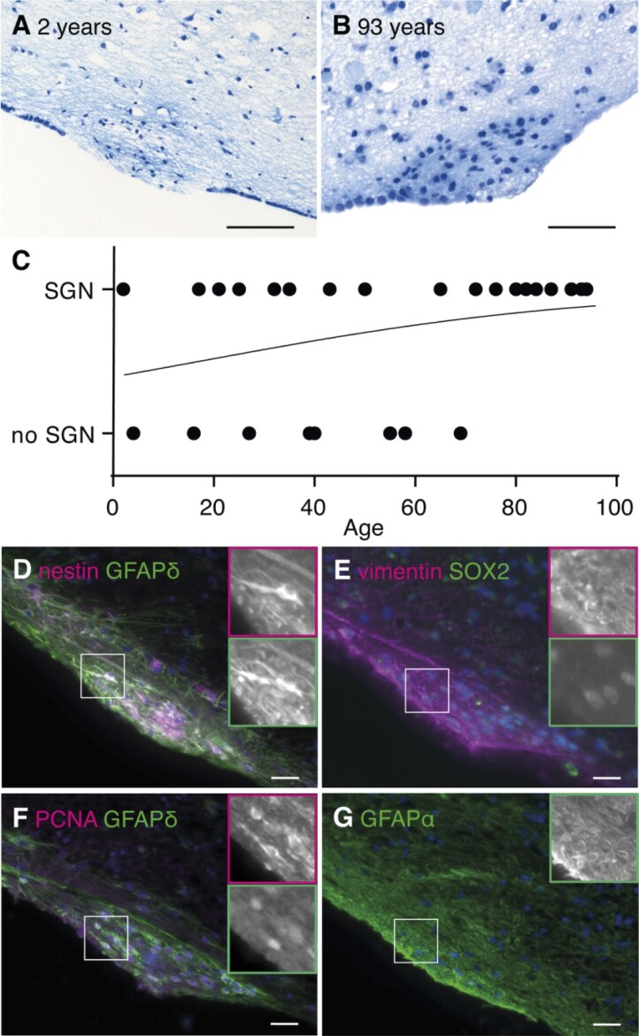
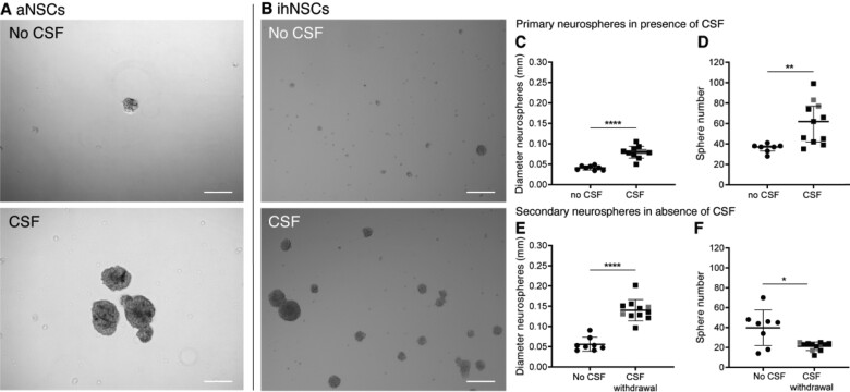
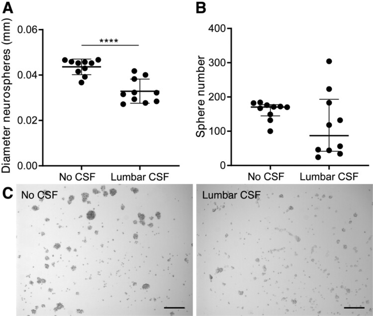
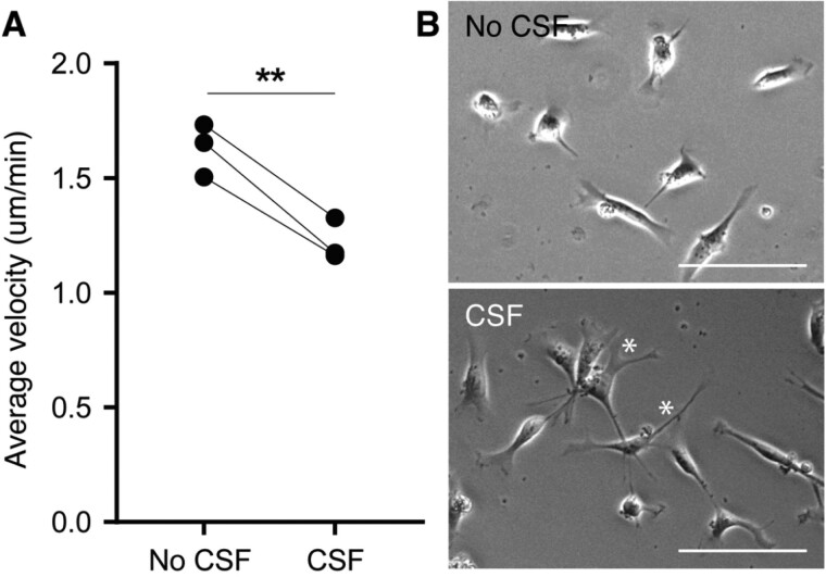
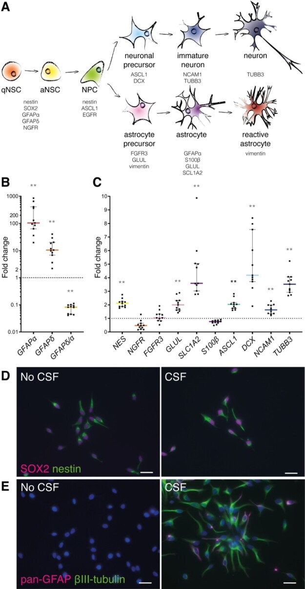
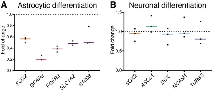
Neurogenesis continues throughout adulthood in specialized regions of the brain. One of these regions is the subventricular zone. During brain development, neurogenesis is regulated by a complex interplay of intrinsic and extrinsic cues that control stem-cell survival, renewal and cell lineage specification. Cerebrospinal fluid (CSF) is an integral part of the neurogenic niche in development as it is in direct contact with radial glial cells, and it is important in regulating proliferation and migration. Yet, the effect of CSF on neural stem cells in the subventricular zone of the adult human brain is unknown. We hypothesized a persistent stimulating effect of ventricular CSF on neural stem cells in adulthood, based on the literature, describing bulging accumulations of subventricular cells where CSF is in direct contact with the subventricular zone. Here, we show by immunohistochemistry on post-mortem adult human subventricular zone sections that neural stem cells are in close contact with CSF via protrusions through both intact and incomplete ependymal layers. We are the first to systematically quantify subventricular glial nodules denuded of ependyma and consisting of proliferating neural stem and progenitor cells, and showed that they are present from foetal age until adulthood. Neurosphere, cell motility and differentiation assays as well as analyses of RNA expression were used to assess the effects of CSF of adult humans on primary neural stem cells and a human immortalized neural stem cell line. We show that human ventricular CSF increases proliferation and decreases motility of neural stem cells. Our results also indicate that adult CSF pushes neural stem cells from a relative quiescent to a more active state and promotes neuronal over astrocytic lineage differentiation. Thus, CSF continues to stimulate neural stem cells throughout aging.

Keywords: cerebrospinal fluid; glial nodules; human; neural stem cells; subventricular zone.


Figures

Graphical Abstract
Graphical Abstract
Figure 1
Figure 1
Contact of neural stem cells with cerebrospinal fluid through protrusions and due to the absence of ependyma covering the subventricular zone (SVZ) of adult donors. (A, F) White dashed lines in two 1 cm thick formalin-fixed human brain slices indicate the three dissected SVZ areas. SVZ1 was dissected in the brain slice containing the most frontal part of the caudate nucleus (cn) and SVZ2 was dissected from the same brain slice, or 1 slice more caudal, under the cingulate gyrus (cg), including part of the corpus callosum (cc). SVZ3 was dissected from the most posterior part of the lateral ventricle (LV). (B, G) Immunofluorescent double labelling of a fresh frozen SVZ tissue section of a control donor (NBB 2005-073, male, 87 years) for GFAPδ (green) and nestin (magenta). White rectangles and asterisks indicate protrusion(s) of NSCs towards the ventricle, of which magnifications are shown in the different channels (C–E and H–J). Nuclei are counterstained with Hoechst. (K) Example of thionin staining showing ependymal cell coverage and absence (transition indicated by the arrow). (L) Ependymal layer coverage in the SVZ of healthy controls is quantified in SVZ1-3 (n =13 donors of SVZ1 and SVZ3, n =14 donors of SVZ2), with significantly less ependymal cell coverage in SVZ2 (one-way ANOVA, F(2,37) = 7.417, P =0.0020; Sidak post-hoc analysis, **P <0.01). Data are expressed as mean ± standard deviation. (M) Sex does not correlate with the average ependymal layer coverage over SVZ1-3 (Mann–Whitney U test, U =13, P =0.1649). Data are expressed as median ± interquartile range. (N) Age does not correlate with the average ependymal layer coverage in this age range (Pearson’s R2 = 0.1601, P =0.1564). Scale bars = 10 μm.
Figure 2
Figure 2
Subventricular glial nodules are commonly present in the human subventricular zone and express proliferation and stem cell markers. (A, B) SGN in the lateral ventricle of a 2-year-old male donor (NBB 1997-017, scale bar 100 μm) and a 93-year-old female donor (NBB 2005-063, scale bar 50 μm), being denuded of ependyma, hypercellular and showing accumulation of thionin-positive fibrous tissue. (C) No significant correlation was found between age and SGN occurrence (Cox–Snell’s R2 = 0.1004, χ2(1) = 2.750, P =0.0972). (D–F) SGNs express NSC (nestin, GFAPδ and SOX2), astrocyte (vimentin) and proliferation (PCNA) markers. (G) GFAPα is not specifically expressed by SGNs (NBB 2005-073, male, 87 years). Scale bars C–F = 10 μm.
Figure 3
Figure 3
Increased neural stem cell (NSC) proliferation upon treatment with ventricular CSF. (A) Representative image of primary adult NSC (aNSC) culture of donor NBB 2012-066. aNSCs treated with 25% pooled post-mortem ventricular CSF were observed to form more and larger neurospheres than untreated aNSCs after 5 days in culture. (B) Representative image of immortalized human NSC (ihNSC) culture without and with CSF of donor NBB 2011-096. ihNSCs treated with 25% post-mortem ventricular CSF (n =11 wells treated with CSF of individual donors) form larger and more neurospheres compared to untreated cells (n =8 untreated wells) after 5 days. (C, D) The ihNSC neurosphere diameter (two-tailed t-test with Welch’s correction, t13.39 = 8.13, P <0.0001) and sphere number (Mann–Whitney U test, U =6, P =0.0018) were quantified after 5 days of CSF treatment. (E, F) Seven days after CSF withdrawal, passaged ihNSC neurosphere diameter (two-tailed t-test, t17 = 7.877, P <0.0001) and number (two-tailed t-test with Welch’s correction, t7.558 = 2.887, P =0.0216) were quantified. In grey, the data points belonging to the wells treated with CSF of Alzheimer patients are indicated; exclusion of these data points did not change the statistical results. Data expressed as mean ± standard deviation in C, E and F, and as median ± interquartile range in D; *P <0.05, **P <0.01, ****P <0.0001; Scale bars = 100 μm.
Figure 4
Figure 4
Decreased NSC proliferation upon treatment with lumbar CSF. (A) Immortalized human NSCs (ihNSCs) treated with 25% lumbar CSF (n =10 wells treated with CSF of individual healthy controls) form smaller neurospheres compared to untreated cells (n =10 untreated wells) after 5 days (two-tailed t-test, t18 = 5.364, P <0.0001). Data are expressed as mean ± standard deviation; ****P <0.0001. (B) ihNSCs tend to form less neurospheres when exposed to lumbar CSF (Mann–Whitney U test, U =31.50, P =0.1717). Data are expressed as median ± interquartile range. (C) After 5 days of treatment with lumbar CSF, ihNSCs adhere more to the bottom of the well and form smaller and less neurospheres compared to untreated ihNSCs. Representative image of neurosphere culture of with CSF of donor G022. Scale bars = 100 μm.
Figure 5
Figure 5
Decreased cell motility and changed morphology of ihNSCs after ventricular CSF pre-treatment. (A) Average velocity of cells pre-treated with CSF (n =3 culture replicates) was significantly lower compared to untreated cells (n =3 culture replicates) (paired t-test, t2 = 10.17, P <0.0095). Per-culture replicate, five cells were traced per five locations per condition. Data are expressed as mean ± standard deviation. (B) Representative phase-contrast pictures of untreated ihNSCs and ihNSCs pre-treated with CSF; the former showing rounder cells, the latter showing a more elongated morphology with more protrusions (*). Scale bars = 100 μm.
Figure 6
Figure 6
Upon treatment with ventricular CSF, ihNSCs transition to a more active state with up-regulation of astrocyte and neuronal progenitor markers. (A) Schematic overview of markers used to characterize the effect of CSF on lineage progression (for references, see Supplementary material Table 5). qNSC = quiescent NSC; aNSC = activated NSC; NPC = neural progenitor cell. (B,C) After 5 days of CSF treatment, cells were collected and used for mRNA expression analysis (n =6 untreated wells, n =11 wells treated with CSF of individual donors); AluS, elongation factor 1-alpha (EF1α) and actin were used as reference genes. GFAPα (Mann–Whitney U test, U =0, P =0.00016), GFAPδ (Mann–Whitney U test, U =0, P =0.00046) and NES (Mann–Whitney U test, U =0, P =0.00016) were higher, whereas the ratio GFAPδ/α (Mann–Whitney U test, U =0, P =0.00046) was lower expressed compared to untreated ihNSCs. NGFR (Mann–Whitney U test, U =11, P =0.0273), FGFR3 (Mann–Whitney U test, U =23, P =0.350) and S100β (Mann–Whitney U test, U =0, P =0.00016) were not differentially expressed, whereas GLUL (Mann–Whitney U test, U =0, P =0.00016) and SLC1A2 (Mann–Whitney U test, U =0, P =0.00016) were higher expressed. ASCL1 (Mann–Whitney U test, U =0, P =0.00016), DCX (Mann–Whitney U test, U =2, P =0.000646) and NCAM1 (Mann–Whitney U test, U =2, P =0.000646) as well as TUBB3 (Mann–Whitney U test, U =0, P =0.00016) were highly expressed compared to untreated ihNSCs. Exclusion of data points belonging to the assays treated with CSF of Alzheimer patients did not change the statistical results. Data are expressed as median fold change of relative mRNA expression of CSF-treated ihNSCs over relative expression of untreated ihNSCs (dotted line) ± interquartile range; Sidak corrected P value = 0.0039 for multiple comparisons, *P <0.0039, **P <0.001, ***P <0.0001, ****P <0.00001. (D) ihNSCs were treated with 25% pooled CSF in ihNSC medium for 5 days and labelled for SOX2 (magenta) and nestin (green). (E) CSF-treated ihNSCs were also stained for the astrocyte marker pan-GFAP (magenta) together with βIII-tubulin (green). Hoechst (blue) as nuclear counter staining. Scale bars = 10 μm.
Figure 7
Figure 7
Ventricular CSF exposure (7 days) primes ihNSCs to differentiate into neuronal over astrocytic lineage upon CSF withdrawal. (A) CSF-treated ihNSCs were differentiated according to astrocytic differentiation protocol upon CSF withdrawal (culture replicates, n =3). Per-culture replicate, ihNSCs were untreated, respectively, pre-treated with pooled CSF of healthy controls. After 7 days, mRNA levels of SOX2 (Mann–Whitney U test, U =0, P =0.1000), GFAPα (Mann–Whitney U test, U =0, P =0.2000), FGFR3 (Mann–Whitney U test, U =0, P =0.1000), SLC1A2 (Mann–Whitney U test, U =0, P =0.1000), S100β (Mann–Whitney U test, U =1, P =0.2000) were measured. (B) Seven days later, ihNSCs were allowed to differentiate according to neuronal differentiation protocol (culture replicates, n =3), after pre-treatment with pooled CSF, mRNA levels of SOX2 (Mann–Whitney U test, U =3, P =0.7000), ASCL1 (Mann–Whitney U test, U =1, P =0.2000), DCX (Mann–Whitney U test, U =3, P =0.7000), NCAM1 (Mann–Whitney U test, U =4, P >0.9999) and TUBB3 (Mann–Whitney U test, U =4, P >0.9999) were measured. Per-culture replicate, ihNSCs were untreated, respectively, pre-treated with pooled CSF of healthy controls. Actin, AluS and glyceraldehyde-3-phosphate dehydrogenase (GAPDH) were used as reference genes. Data are expressed as median fold change of relative mRNA expression of pre-treated ihNSCs over relative expression of untreated ihNSCs (dotted line); Sidak corrected P value = 0.0102.

Similar articles

Cited by

References

    1. Adams CW, Abdulla YH, Torres EM, Poston RN. Periventricular lesions in multiple sclerosis: their perivenous origin and relationship to granular ependymitis. Neuropathol Appl Neurobiol 1987; 13: 141–52. - PubMed
    1. Alonso MI, Lamus F, Carnicero E, Moro JA, de la Mano A, Fernández JMF, et al.Embryonic cerebrospinal fluid increases neurogenic activity in the brain ventricular-subventricular zone of adult mice. Front Neuroanat 2017; 11: 124. - PMC - PubMed
    1. Andersen J, Urbán N, Achimastou A, Ito A, Simic M, Ullom K, et al.A transcriptional mechanism integrating inputs from extracellular signals to activate hippocampal stem cells. Neuron 2014; 83: 1085–97. - PMC - PubMed
    1. Baird GS, Nelson SK, Keeney TR, Stewart A, Williams S, Kraemer S, et al.Age-dependent changes in the cerebrospinal fluid proteome by slow off-rate modified aptamer array. Am J Pathol 2012; 180: 446–56. - PMC - PubMed
    1. Basak O, Giachino C, Fiorini E, Macdonald HR, Taylor V. Neurogenic subventricular zone stem/progenitor cells are Notch1-dependent in their active but not quiescent state. J Neurosci 2012; 32: 5654–66. - PMC - PubMed

LinkOut - more resources