. 2020 Nov 27;2(2):fcaa203.
doi: 10.1093/braincomms/fcaa203. eCollection 2020.

Transcranial magnetic stimulation reveals diminished homoeostatic metaplasticity in cognitively impaired adults

Affiliations

Transcranial magnetic stimulation reveals diminished homoeostatic metaplasticity in cognitively impaired adults

Mark H Sundman et al. Brain Commun. .

Abstract

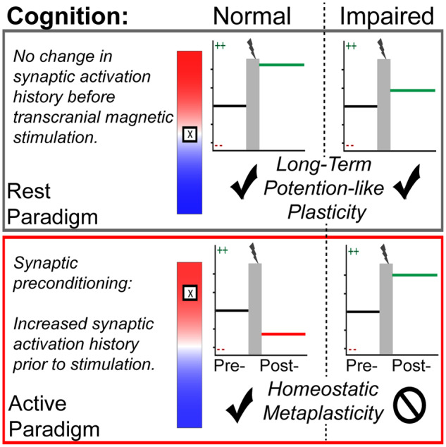
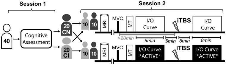
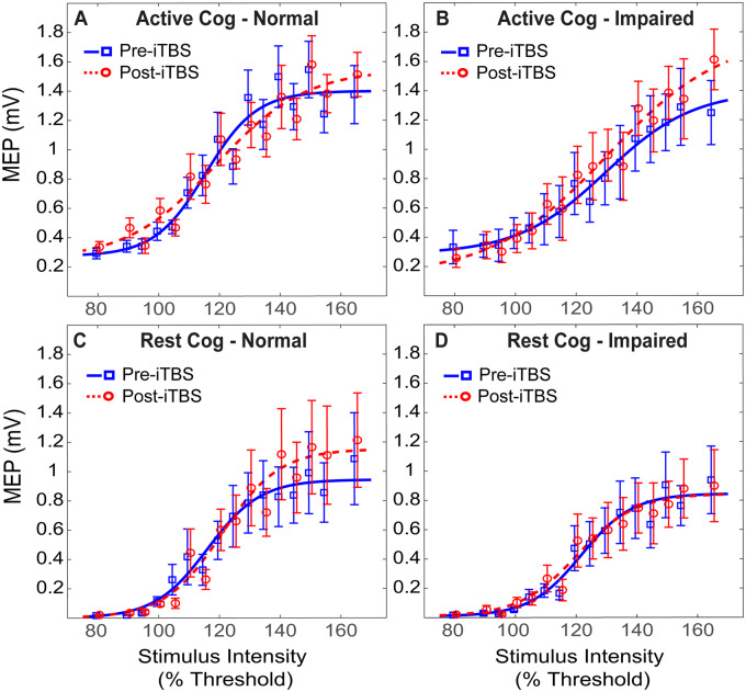
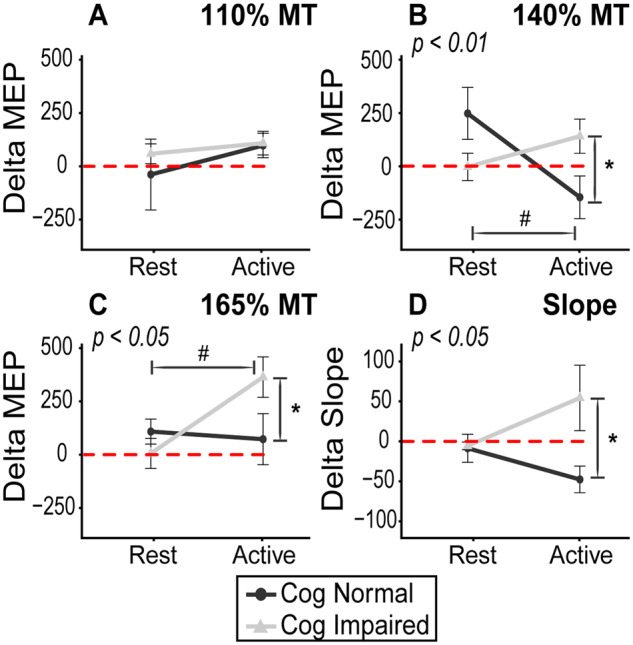
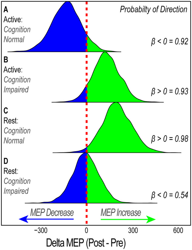
Homoeostatic metaplasticity is a neuroprotective physiological feature that counterbalances Hebbian forms of plasticity to prevent network destabilization and hyperexcitability. Recent animal models highlight dysfunctional homoeostatic metaplasticity in the pathogenesis of Alzheimer's disease. However, the association between homoeostatic metaplasticity and cognitive status has not been systematically characterized in either demented or non-demented human populations, and the potential value of homoeostatic metaplasticity as an early biomarker of cognitive impairment has not been explored in humans. Here, we report that, through pre-conditioning the synaptic activity prior to non-invasive brain stimulation, the association between homoeostatic metaplasticity and cognitive status could be established in a population of non-demented human subjects (older adults across cognitive spectrums; all within the non-demented range). All participants (n = 40; age range, 65-74, 47.5% female) underwent a standardized neuropsychological battery, magnetic resonance imaging and a transcranial magnetic stimulation protocol. Specifically, we sampled motor-evoked potentials with an input/output curve immediately before and after repetitive transcranial magnetic stimulation to assess neural plasticity with two experimental paradigms: one with voluntary muscle contraction (i.e. modulated synaptic activity history) to deliberately introduce homoeostatic interference, and one without to serve as a control condition. From comparing neuroplastic responses across these experimental paradigms and across cohorts grouped by cognitive status, we found that (i) homoeostatic metaplasticity is diminished in our cohort of cognitively impaired older adults and (ii) this neuroprotective feature remains intact in cognitively normal participants. This novel finding suggests that (i) future studies should expand their scope beyond just Hebbian forms of plasticity that are traditionally assessed when using non-invasive brain stimulation to investigate cognitive ageing and (ii) the potential value of homoeostatic metaplasticity in serving as a biomarker for cognitive impairment should be further explored.

Keywords: TMS; cognitive ageing; dementia; mild cognitive impairment; plasticity.


Figures

Graphical Abstract
Graphical Abstract
Figure 1
Figure 1
Experimental design. Participants were stratified by cognitive status and randomly assigned to participate in one of the two experimental paradigms. Thus, there were four experimental cohorts, each with 10 participants. CN: cognitively normal; CI: cognitively impaired; MRI: magnetic resonance imaging; MVC: maximum voluntary contraction; MT: motor threshold; I/O: input/output; iTBS: intermittent theta burst stimulation
Figure 2
Figure 2
Input–output curve. (A) A repetitive I/O curve during the rest paradigm. We examined the neuroplastic response at four distinct components of the curve: (I)110% MT, (II)140% MT, (III) 165% MT and (IV) the calculate slope of the fitted curve. (B) An example of MEP waveform. In this example, the TMS intensity was 145% MT and the peak-to-peak amplitude was determined to be ∼2100 uV. For each curve, 64 such MEPs were plotted with amplitude as a function of TMS intensity. The sigmoidal curve is fit with the Boltzmann equation
Figure 3
Figure 3
Raw MEPs Pre- and Post-iTBS. The averaged pre- and post-iTBS I/O curves are plotted for each cohort. The mean MEPs for each intensity are plotted at both time points, and the sigmoidal I/O curve is fit with the Boltzmann equation. Blue denotes baseline cortical excitability profiles pre-iTBS and red denotes post-iTBS cortical excitability profiles. The difference between the two reflects response to iTBS, in which an LTP-like effect is observed when post-iTBS MEP amplitudes (red) are greater than baseline MEP values (blue)
Figure 4
Figure 4
Interaction plots on normalized MEP data. The interaction effect of cognitive status and experimental paradigm on normalized MEP values are shown. Values > 0 represent an excitatory (LTP-like) iTBS effect and, and values < 0 reflect an inhibitory (long-term depression-like) response. This plot illustrates the divergent responses to iTBS by cognition and paradigm. There is a greater response to iTBS in the active paradigm in the cognitively impaired participants, but the opposite is true among cognitively normal participants. This is most evident 140% MT (B) where there is predominant late I-wave contribution, which is known to underly iTBS effects. The P-values depicted in subtitles indicate interaction effect result from two-way ANOVA (GROUP × PARADIGM). As indicated by FLSD, * denotes pairwise significant differences in group responses (within paradigm) and # denotes pairwise significant differences between conditions (within group)
Figure 5
Figure 5
Posterior distribution for possible parameter values at 140% of MT. Derived from the Bayesian analysis of normalized MEP values at 140% MT, this figure shows the probability distribution of possible parameter values for each experimental cohort. The probability of direction value represents the probability mass of the posterior distribution that falls above or below zero, representing a predicted increase or decrease, respectively, in MEP values. Values of 0.5 indicate 50% chance of increase or decrease, whereas value of 1.0 indicates 100% probability of a specified direction. This figure shows that iTBS results in: (A) 92% probability that MEP will decrease in cognitively normal individuals when preceding synaptic activation history is high (apparent homoeostatic interference), (B) 93% probability that MEP will increase cognitively impaired individuals when preceding synaptic activation history is high (diminished homoeostatic metaplasticity), (C) 98% MEP will increase in cognitively normal individuals when preceding synaptic activation history is unaltered (strong LTP-like response), (D) ∼50% probability that MEP will either increase or decrease (weaker LTP-like response) in cognitively impaired individuals when preceding synaptic activation history is unaltered

Similar articles

Cited by

References

    1. Abbott LF, Nelson SB. Synaptic plasticity: taming the beast. Nat Neurosci 2000; 3: 1178–83. - PubMed
    1. Abraham WC. Metaplasticity: tuning synapses and networks for plasticity. Nat Rev Neurosci 2008; 9: 387–99. - PubMed
    1. Abraham WC, Bear MF. Metaplasticity : plasticity of synaptic. Trends Neurosci 1996; 19: 126–30. - PubMed
    1. Abraham WC, Robins A. Memory retention—the synaptic stability versus plasticity dilemma. Trends Neurosci 2005; 28: 73–8. - PubMed
    1. Amatniek JC, Hauser WA, DelCastillo-Castaneda C, Jacobs DM, Marder K, Bell K, et al. Incidence and predictors of seizures in patients with Alzheimer’s disease. Epilepsia 2006; 47: 867–72. - PubMed

LinkOut - more resources