

doi: 10.1016/j.xpro.2020.100146.
eCollection 2020 Dec 18.
Protocol for Patch-Seq of Small Interneurons
Affiliations
- PMID: 33377040
- PMCID: PMC7757288
- DOI: 10.1016/j.xpro.2020.100146
Item in Clipboard
Protocol for Patch-Seq of Small Interneurons
STAR Protoc.
.
Abstract
Obtaining electrophysiological recordings and gene expression information from the same neuron (Patch-seq) brings forward a unique opportunity to study the transcriptional correlates of functional properties and vice versa. Here, we provide a detailed Patch-seq protocol tailored to the specialized demands of studying small interneurons. Focusing on the technically demanding process of transitioning between patch recordings and cell extraction, our protocol describes and troubleshoots steps for successfully collecting small interneurons, allowing for multi-modal Patch-seq interrogation of this crucial cell type.
© 2020 The Author(s).
Conflict of interest statement
The authors declare no competing interests.
Figures


Equipment for Cell Collection (A) Mouth pipetting assembly used for expelling the collected cell into the collection tube. (B) Metal plate sitting on dry ice used for flash-freezing samples.

Schematic Timeline for Patch Recordings and Cell Extraction Corresponding representative seal test protocols are shown for each step. Step 1: A clean, unobstructed pipette is lowered into the vicinity of the target cell. Step 2: A giga-ohm seal is formed between the pipette and cell. Step 3: The cell membrane is ruptured yielding electrical access to the inside of the cell. At this point the membrane properties of the cell can be interrogated (e.g., inset trace, showing cell response to 500 ms of depolarizing current injected in current-clamp mode). Step 4: Negative pressure is applied, drawing the cell contents into the pipette and forming a strong seal onto the cell nucleus. Step 5: The pipette is slowly retracted from the tissue bringing the cell nucleus with it. Seal test recordings shown here were obtained by holding the cell at −60 mV and stepping the membrane voltage sequentially to −50 mV and then −70 mV for 10 ms before returning to the holding potential. Scale bar, 10 ms and 1 nA for #1; 10 ms and 200 pA for #2–5; 100 ms and 25 mV for inset in #3.

Scoring System for Assessing Cell Extraction Perfectly extracted cells must meet all the criteria outlined: successfully extracted nucleus, seal maintained throughout the whole extraction process, no debris observed on the outside of the pipette, no contact of the tip of the pipette with any surface before it reaches the lysis buffer in the collection tube, and less than 10 min duration for the entire extraction process. Assign a score to each cell and only process for sequencing cells with “perfect” or “very good” scores. Cells with “good” scores can also be sequenced if, for example, sample numbers are low or there is particularly interesting physiology.
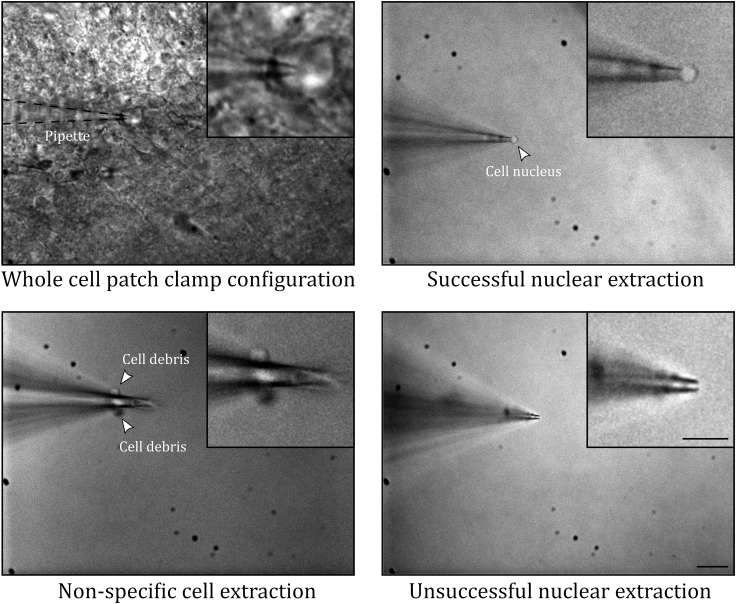
Example Images of Patching and Cell Collection Top left: Whole-cell patch-clamp recording of a glomerular layer interneuron from an acute slice of juvenile mouse olfactory bulb. Top right: Pipette held above the slice after a successful extraction of the cell nucleus. Bottom left: Post-recording pipette with non-specific cellular debris attached to the side of the glass. Bottom right: Post-recording pipette where the nucleus has become dislodged from the pipette tip during the extraction procedure. Scale bar, 20 μm for the main panels and 10 μm for the insets.
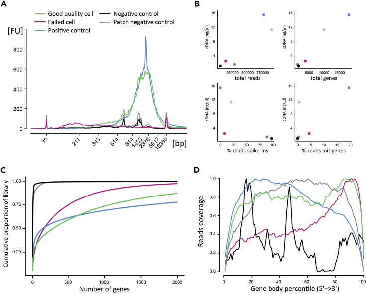
Data from Five Representative Example Patch-Seq-ed Cells, Illustrating Different Potential Outcomes in Quality Control Metrics for cDNA Preparation and scRNA-Seq (A) Bioanalyzer capillary electrophoresis traces. (B) scRNA-seq quality metrics, showing per sample cDNA concentration, total number of mapped reads per cell, total number of genes detected per cell, percentage of reads mapping to technical ERCC spike-ins per cell and percentage of reads mapping to mitochondrial genes per cell. (C) Library complexity depicted as the cumulative proportion of the total number of genes detected per cell. (D) Reads mapping along gene body length showing full-length cDNA coverage for good quality samples. Green, good quality cell; red, low-quality cell; blue, positive control; black, negative control; gray, patch negative control.
References
Publication types
MeSH terms
LinkOut - more resources
Full Text Sources
Molecular Biology Databases
