Diagnostic analysis of vertical orbital dystopia and canthal tilt for surgical correction
- PMID: 33377462
- PMCID: PMC7783177
- DOI: 10.5125/jkaoms.2020.46.6.379
Diagnostic analysis of vertical orbital dystopia and canthal tilt for surgical correction
Abstract
Objectives: We sought to identify a clinically useful method of analyzing orbital dystopia to aid in diagnosis and treatment planning and to quantify vertical discrepancies in eye level and variations in canthal tilt in Koreans.
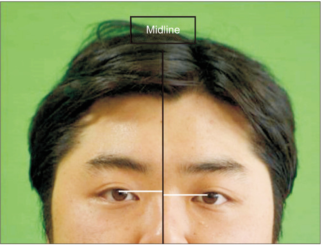
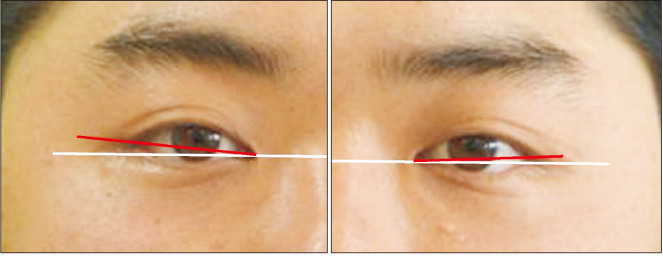
Patients and methods: In 76 Korean patients with a mean age of 23.12 years, mean differences in the level of the pupils, lateral canthi, medial canthi, and canthal tilt were measured. The difference in pupil level was calculated from the perpendicular lines drawn from the midpupil area of each eye to the midline of the face to determine the amount of skeletal discrepancy of the eye. Soft tissue discrepancies were determined according to the vertical difference between the lines drawn from the lateral or medial canthus of each eye perpendicular to the midline of the face. The canthal tilt was determined from the inclination of a line connecting the lateral and medial canthi, then classified as class I, II, or III.
Results: Mean differences in pupil level, medial canthi, and lateral canthi were 1.57±1.10 mm, 1.14±1.07 mm, and 2.03±1.64 mm, respectively. The mean degree of canthal tilt were 8.45°±3.53° for the right side and 8.42°±3.81° for the left side. No study participants presented with class III canthal tilt. The mean canthal tilt values for those with class I tilt were 3.21°±1.68° for the right side and 3.18°±1.63° for the left side, while, for those who had class II tilt, the values were 9.60°±3.66° for the right side and 9.54°±2.99° for the left side.
Conclusion: The presented diagnostic method of orbital dystopia can be used to effectively establish a treatment plan that takes into consideration the patient's skeletal and soft-tissue discrepancies.
Keywords: Anthropometry; Facial asymmetry; Orbit.
Conflict of interest statement
No potential conflict of interest relevant to this article was reported.
Figures


Similar articles
-
Anatomy of the lateral canthal tendon.Oral Surg Oral Med Oral Pathol Oral Radiol Endod. 2000 Jan;89(1):24-8. doi: 10.1016/s1079-2104(00)80009-8. Oral Surg Oral Med Oral Pathol Oral Radiol Endod. 2000. PMID: 10630937
-
Ocular adnexal asymmetry in models: a magazine photograph analysis.Can J Ophthalmol. 2006 Apr;41(2):175-82. doi: 10.1139/I06-005. Can J Ophthalmol. 2006. PMID: 16767204
-
Is medial canthal tilt a powerful cue for facial attractiveness?Ophthalmic Plast Reconstr Surg. 2007 Jan-Feb;23(1):52-6. doi: 10.1097/IOP.0b013e31802dd7dc. Ophthalmic Plast Reconstr Surg. 2007. PMID: 17237692
-
Lateral canthal anatomy: a review.Orbit. 2012 Aug;31(4):279-85. doi: 10.3109/01676830.2012.694957. Epub 2012 Jun 12. Orbit. 2012. PMID: 22690873 Review.
-
Lateral canthal surgery.Facial Plast Surg. 2010 Aug;26(3):193-200. doi: 10.1055/s-0030-1254329. Epub 2010 Jun 3. Facial Plast Surg. 2010. PMID: 20524167 Review.
Cited by
-
Endoscopic-assisted versus open fronto-orbital distraction for unicoronal craniosynostosis: morphometric and technique considerations.Childs Nerv Syst. 2024 Dec 18;41(1):59. doi: 10.1007/s00381-024-06662-8. Childs Nerv Syst. 2024. PMID: 39692920 Free PMC article.
References
-
- Choi KY. Analysis of facial asymmetry. Arch Craniofac Surg. 2015;16:1–10. doi: 10.7181/acfs.2015.16.1.1. https://doi.org/10.7181/acfs.2015.16.1.1 . - DOI - PMC - PubMed
-
- Song WC, Koh KS, Kim SH, Hu KS, Kim HJ, Park JC, et al. Horizontal angular asymmetry of the face in Korean young adults with reference to the eye and mouth. J Oral Maxillofac Surg. 2007;65:2164–8. doi: 10.1016/j.joms.2006.11.018. https://doi.org/10.1016/j.joms.2006.11.018 . - DOI - PubMed
-
- Hwang HS, Youn IS, Lee KH, Lim HJ. Classification of facial asymmetry by cluster analysis. Am J Orthod Dentofacial Orthop. 2007;132:279.e1–6. doi: 10.1016/j.ajodo.2007.01.017. https://doi.org/10.1016/j.ajodo.2007.01.017 . - DOI - PubMed
-
- Altug-Atac AT, Grayson BH, McCarthy JG. Comparison of skeletal and soft-tissue changes following unilateral mandibular distraction osteogenesis. Plast Reconstr Surg. 2008;121:1751–9. doi: 10.1097/PRS.0b013e31816aa003. https://doi.org/10.1097/PRS.0b013e31816aa003 . - DOI - PubMed
-
- Yamashita Y, Nakamura Y, Shimada T, Nomura Y, Hirashita A. Asymmetry of the lips of orthognathic surgery patients. Am J Orthod Dentofacial Orthop. 2009;136:559–63. doi: 10.1016/j.ajodo.2007.10.057. https://doi.org/10.1016/j.ajodo.2007.10.057 . - DOI - PubMed