

Design and Pilot data of the high myopia registration study: Shanghai Child and Adolescent Large-scale Eye Study (SCALE-HM)
- PMID: 33377612
- PMCID: PMC8359463
- DOI: 10.1111/aos.14617
Design and Pilot data of the high myopia registration study: Shanghai Child and Adolescent Large-scale Eye Study (SCALE-HM)
Abstract
Purpose: To describe the methodology and pilot data of the Shanghai Child and Adolescent Large-scale Eye Study (SCALE-HM).
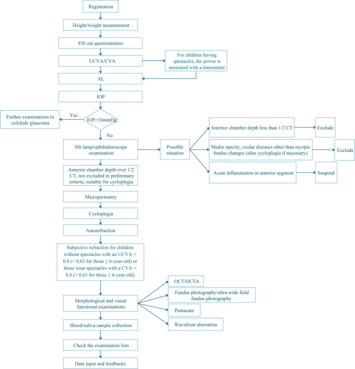
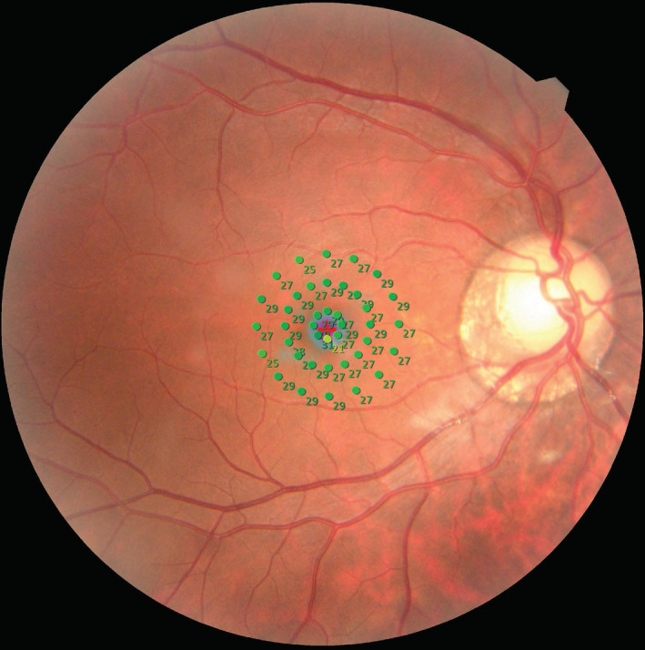
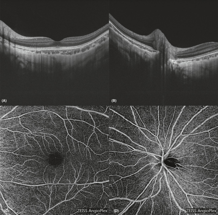
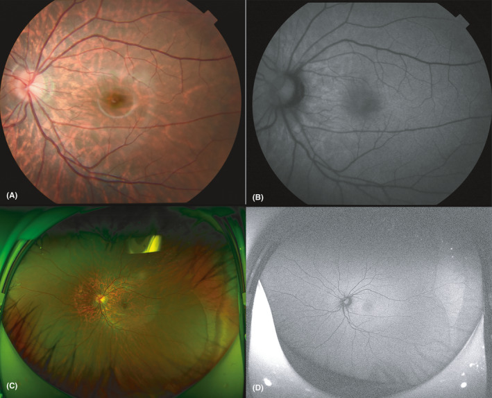
Methods: This is a population-based, prospective, examiner-masked study with annual follow-up. Patients are 4- to 18-year-olds with high myopia. The participants will fill out questionnaires and then undergo visual acuity, axial length (AL), intraocular pressure, ophthalmologist assessment, microperimetry, cycloplegic refraction, Pentacam, wavefront aberration, fundus, blood and saliva examinations. To describe the pilot data, intergroup differences were assessed with t-tests or analysis of variance and a logistic regression model was used to determine the independent factors associated with peripapillary atrophy (PPA).
Results: Overall, 134 eyes of 79 participants met the pilot study recruitment criteria. The mean AL and spherical equivalent were 26.91 ± 1.07 mm and -9.40 ± 1.77 D, respectively. Peripapillary atrophy (PPA) (N = 112) and tessellated fundus (N = 67) were the most common fundus changes. The mean AL was significantly longer in PPA (27.08 ± 0.93 mm) than in non-PPA eyes (26.06 ± 1.31 mm; p < 0.001). Axial length (AL) (p = 0.041) was the only independent factor associated with PPA. Axial length (AL) was significantly longer in eyes with diffuse chorioretinal atrophy (N = 11; 28.02 ± 1.31 mm) than without myopic retinal lesions (N = 56; 26.48 ± 0.91 mm, p < 0.001) or with tessellated fundus (N = 67; 27.09 ± 0.97 mm, p = 0.012). The myopic degree was higher in eyes with diffuse chorioretinal atrophy than without myopic retinal lesions (-10.51 ± 2.76 D versus -9.06 ± 1.58 D, p = 0.039).
Conclusion: Peripapillary atrophy and tessellated fundus were common in children and adolescents with high myopia. Results from this prospective study will help to understand the mechanisms, development and prognosis of these changes and can guide early myopia screening.
Keywords: children; fundus change; high myopia; registration study.
© 2020 The Authors. Acta Ophthalmologica published by John Wiley & Sons Ltd on behalf of Acta Ophthalmologica Scandinavica Foundation.
Figures
References
-
- Achenbach TM & Ruffle TM (2000): The Child Behavior Checklist and related forms for assessing behavioral/emotional problems and competencies. Pediatr Rev 21: 265–271. - PubMed
-
- Al‐Sheikh M, Phasukkijwatana N, Dolz‐Marco R, Rahimi M, Iafe NA, Freund KB, Sadda SR & Sarraf D (2017): Quantitative OCT angiography of the retinal microvasculature and the Choriocapillaris in myopic eyes. Invest Ophthalmol Vis Sci 58: 2063–2069. - PubMed
-
- Alzaben Z, Cardona G, Zapata MA & Zaben A (2017): Interocular asymmetry in choroidal thickness and retinal sensitivity in high myopia. Retina 38: 1620–1628. - PubMed
-
- Byer NE (1965): Clinical study of lattice degeneration of the retina. Trans Am Acad Ophthalmol Otolaryngol 69: 1065–1081. - PubMed
Publication types
MeSH terms
Grants and funding
- 2017YQ019/Municipal Human Resources Development Program for Outstanding Young Talents in Medical and Health Sciences in Shanghai
- 81900911/National Natural Science Foundation of China
- 2017ZX09304010/National Science and Technology Major Project of China
- 18YF1420200/Shanghai Sailing Program
- 2016YFC0904800/National Key R&D Program of China
LinkOut - more resources
Full Text Sources
Medical
Miscellaneous
