. 2020 Dec;10(8):e263.
doi: 10.1002/ctm2.263.

MiRNA-516a promotes bladder cancer metastasis by inhibiting MMP9 protein degradation via the AKT/FOXO3A/SMURF1 axis

Affiliations

MiRNA-516a promotes bladder cancer metastasis by inhibiting MMP9 protein degradation via the AKT/FOXO3A/SMURF1 axis

Yuanyuan Chang et al. Clin Transl Med. 2020 Dec.

Abstract

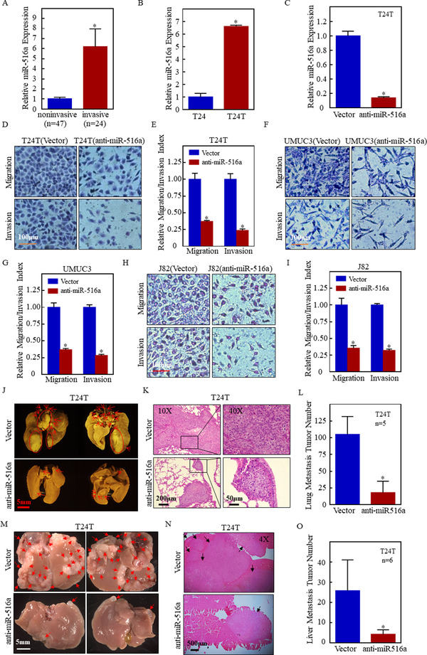
Background: Metastasis is the leading cause of death in patients with bladder cancer (BC). However, current available treatments exert little effects on metastatic BC. Moreover, traditional grading and staging have only a limited ability to identify metastatic BC. Accumulating evidence indicates that the aberrant expression of microRNA is intimately associated with tumor progression. So far, many miRNAs have been identified as molecular targets for cancer diagnosis and therapy. This study focused on the role of miR-516a-5p (miR-516a) in BC.

Methods: MiR-516a expression and its downstream signaling pathway were detected using molecular cell biology and biochemistry approaches and techniques. Fresh clinical BC tissue was used to study the clinicopathological characteristics of patients with different miR-516a expression. The biological functions of miR-516a in BC were tested both in vivo and in vitro.

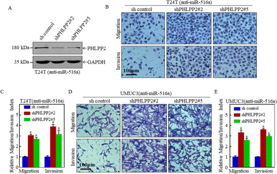
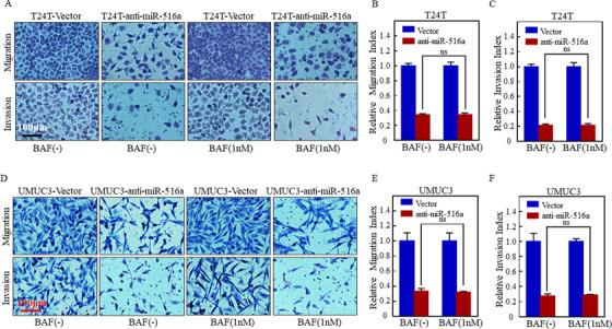
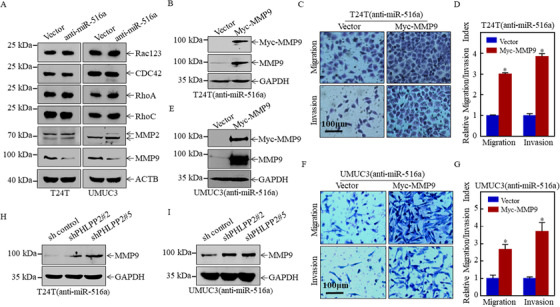
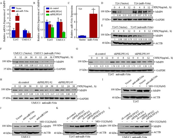
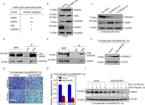
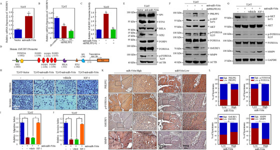
Results: A more invasive BC phenotype was significantly and positively correlated with miR-516a overexpression in BC patients. MiR-516a inhibition significantly decreased BC cell invasion and migration in vitro and in vivo. Furthermore, miR-516a attenuated the expression of PH domain leucine-rich repeat-containing protein phosphatase 2 protein and inhibited SMAD-specific E3 ubiquitin protein ligase 1 transcription by activating the AKT/Forkhead box O3 signaling pathway, which stabilized MMP9 and slowed down its proteasomal degradation, ultimately promoting BC motility and invasiveness.

Conclusions: Our findings reveal the crucial function of miR-516a in promoting BC metastasis, and elucidate the molecular mechanism involved, suggesting that miR-516a may be a promising novel diagnostic and therapeutic target for BC.

Keywords: PHLPP2; bladder cancer; metastasis; miR-516a; migration and invasion.


Conflict of interest statement

The authors declare no conflict of interest.

Figures

FIGURE 1
FIGURE 1
Suppression of miR‐516a inhibited BC cell metastasis in vitro and in vivo. (A) Real‐time PCR was performed to detect miR‐516a expression in human‐invasive BC tissues and noninvasive BC tissues. (B) Real‐time PCR was performed to detect miR‐516a expression in T24 and T24T cell lines. (C) Inhibitory efficiency of miR‐516ain T24T cells was verified by real‐time PCR. (D, F, and H) Transwell assay performed in transfected T24T/UMUC3/J82 cells to evaluate cell migration and invasion ability. Scale bar: 100 μm. (E, G, and I) Graphical representation of panels D, F, and H and statistical analysis. (J‐L) T24T (vector) and T24T (anti‐miR‐516a) cells were injected into nude mice through the tail vein. Images of nude mouse lungs taken using Zeiss SteREO Discovery v.20 (J). (K) Metastatic colonization was confirmed using H&E staining. (L) Lung metastases were counted after post‐fixation with neutral‐buffered formalin/Bouin's solution fixative. The histogram indicates mean ± SD of the number of lung metastases from five mice in each group. (M‐O) T24T (vector) and T24T (anti‐miR‐516a) cells were injected into nude mice through the spleen. Zeiss SteREO Discovery v.20 was used to take the images of nude mice livers (M). (N) Metastatic colonization was confirmed using H&E staining. (O) Liver metastases were counted after post‐fixation with 4% PFA. The histogram indicates mean ± SD of the number of liver metastases from six mice in each group
FIGURE 2
FIGURE 2
PHLPP2 knockdown plays an essential role in miR‐516a‐induced migration and invasion of BC cells. (A) Knockdown efficiency of PHLPP2 was confirmed in T24T (anti‐miR‐516a) cells by western blot. (B and D) Transwell assay in indicated cells to evaluate the role of PHLPP2 in miR‐516a‐promoted BC cell migration and invasion. Scale bar: 100 μm. (C and E) Graphical representation of panels B and D and statistical analysis
FIGURE 3
FIGURE 3
BAF did not affect the promotion of migration and invasion exerted by miR‐516a. (A and D) Transwell assay was performed in transfected T24T/UMUC3 cells treated with or without BAF (1 nM) to evaluate cell migration and invasion. Scale bar: 100 μm. (B, C, E, and F) ns indicates no significant difference between the vehicle‐ and BAF‐treated groups (< .05)
FIGURE 4
FIGURE 4
MMP9 played a major role in miR‐516a‐mediated promotion of BC metastasis. (A) Effect of miR‐516a on the protein expression of Rac123, CDC42, RhoA, RhoC, MMP2, and MMP9 in T24T and UMUC3 cells by western blotting. Cells were seeded at the specified densities and starved in 0.1% FBS DMEM or 1:1 (DMEM:F12) for 12 h. Subsequently, the cells were incubated in 10% FBS DMEM or 5% 1:1 (DMEM: F12) for an additional 12 h, and then the cell lysates were subjected to western blotting. (B and E) Overexpression of Myc‐MMP9 in T24T/UMUC3 (anti‐miR‐516a) cells was confirmed by western blot. (C, D, F, and G) Transwell assay was performed in transfected T24T/UMUC3 (anti‐miR‐516a) cells after ectopic expression of Myc‐MMP9. (H and I) The cells indicated were lysed and subjected to western blotting to monitor MMP9 protein expression
FIGURE 5
FIGURE 5
MiR‐516a stabilized MMP9 by inhibiting its degradation by the proteasome. (A) MMP9 mRNA expression in T24T/UMUC3 (vector) and T24T/UMUC3 (anti‐miR‐516a) cells, as determined by real‐time PCR. (B) MMP9 mRNA expression in cells indicated, as determined by real‐time PCR. (C) Real‐time PCR was performed to detect miR‐516a expression after ectopic expression of miR‐516a in T24 cells. (D) Rate of MMP9 degradation in T24 (vector) and T24 (miR‐516a) cells, as determined by western blotting. (E and F) T24T/UMUC3 (vector) and T24T/UMUC3 (anti‐miR‐516a) cells were treated with CHX (50 μg/mL) and harvested at the indicated times. The cell lysate was subjected to western blotting with the indicated antibodies. (G and H) Rate of MMP9 degradation in T24T/UMUC3 (anti‐miR‐516a/sh control), T24T/UMUC3 (anti‐miR‐516a/shPHLPP2 #2), and T24T/UMUC3 (anti‐miR‐516a/shPHLPP2 #5) cells, as determined by western blotting. (I‐L) Cells were treated with MG‐132 for 10 h before harvest; protein accumulation was observed by western blotting
FIGURE 6
FIGURE 6
SMURF1 bound MMP9 to promote its degradation, thereby inhibiting BC migration and invasion. (A) Mass spectrometric analysis was performed to identify MMP9‐associated proteins. (B and C) Expression of HSPA8, CBL, SMURF1, and MMP9 in the indicated cell extracts, as determined by western blotting. (D and E) Immunoprecipitation was performed on 293T cell lysates with control IgG (D), anti‐Myc, or (E) anti‐FLAG antibody. The immunoprecipitate was assayed for MMP9 and SMURF1 levels. (F) T24T (anti‐miR‐516a/shPHLPP2 #2/vector) cells and T24T (anti‐miR‐516a/shPHLPP2 #2/Flag‐SMURF1) cells were lysed, and overexpression efficiency of SMURF1 was evaluated by western blotting. (G and H) Invasion and migration of T24T (anti‐miR‐516a/shPHLPP2 #2/vector) and T24T (anti‐miR‐516a/shPHLPP2 #2/Flag‐SMURF1) cells were evaluated by transwell assay. Data were expressed as mean ± SD, and the asterisk (*) indicates a significant difference. (I) The degradation rate of MMP9 was monitored in T24T (anti‐miR‐516a/shPHLPP2 #2/vector) and T24T (anti‐miR‐516a/shPHLPP2 #2/Flag‐SMURF1) cells by western blotting. ImageJ was used to quantify MMP9 expression relative to ACTB
FIGURE 7
FIGURE 7
MiR‐516a/PHLPP2 promoted BC migration and invasion via AKT/FOXO3A/SMURF1 signaling pathway. (A and B) SMURF1 mRNA expression, as determined by real‐time PCR. (C) Luciferase activity was evaluated after SMURF1 promoter plasmid and pRL‐TK were transiently co‐transfected into the indicated cells. (D) Bioinformatics software was used to predict transcription factor binding sites of human SMURF1 promoter. (E‐G) The indicated proteins were analyzed by western blotting. (H‐J) Transwell assay was performed in T24T (Vector), T24T (anti‐miR‐516a), T24T (anti‐miR‐516a, vehicle) and T24T (anti‐miR‐516a, IGF‐1) cells. (K) Immunohistochemical staining of PHLPP2, p‐FOXO3A, SMURF1, and MMP9 in 71 BC clinical specimens; 100× magnification, scale bar: 200 μm; 400× magnification, scale bar: 50 μm. (L) Correlation between miR‐516a and PHLPP2/p‐FOXO3A/SMURF1/MMP9 in 71 clinical BC specimens

References

    1. Siegel RL, Miller KD, Jemal A. Cancer statistics, 2019. CA Cancer J Clin. 2019;69(1):7‐34. - PubMed
    1. Siegel RL, Miller KD, Jemal A. Cancer statistics, 2020. CA Cancer J Clin. 2020;70(1):7‐30. - PubMed
    1. Miller KD, Nogueira L, Mariotto AB, Rowland JH, Siegel RL. Cancer treatment and survivorship statistics, 2019. CA Cancer J Clin. 2019;69(5):363‐385. - PubMed
    1. Chamie K, Litwin MS, Bassett JC, et al. Recurrence of high‐risk bladder cancer: a population‐based analysis. Cancer. 2013;119(17):3219‐3227. - PMC - PubMed
    1. Vishnu P, Mathew J, Tan W. Current therapeutic strategies for invasive and metastatic bladder cancer. Onco Targets Ther. 2011;4:97‐113. - PMC - PubMed