

Decreasing brain iron in multiple sclerosis: The difference between concentration and content in iron MRI
- PMID: 33378095
- PMCID: PMC7927296
- DOI: 10.1002/hbm.25306
Decreasing brain iron in multiple sclerosis: The difference between concentration and content in iron MRI
Abstract
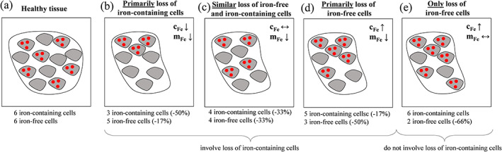
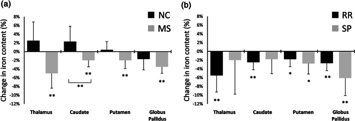
Increased brain iron concentration is often reported concurrently with disease development in multiple sclerosis (MS) and other neurodegenerative diseases. However, it is unclear whether the higher iron concentration in patients stems from an influx of iron into the tissue or a relative reduction in tissue compartments without much iron. By taking into account structural volume, we investigated tissue iron content in the deep gray matter (DGM) over 2 years, and compared findings to previously reported changes in iron concentration. 120 MS patients and 40 age- and sex-matched healthy controls were included. Clinical testing and MRI were performed both at baseline and after 2 years. Overall, iron content was calculated from structural MRI and quantitative susceptibility mapping in the thalamus, caudate, putamen, and globus pallidus. MS patients had significantly lower iron content than controls in the thalamus, with progressive MS patients demonstrating lower iron content than relapsing-remitting patients. Over 2 years, iron content decreased in the DGM of patients with MS, while it tended to increase or remain stable among controls. In the thalamus, decreasing iron content over 2 years was associated with disability progression. Our study showed that temporally increasing magnetic susceptibility in MS should not be considered as evidence for iron influx because it may be explained, at least partially, by disease-related atrophy. Declining DGM iron content suggests that, contrary to the current understanding, iron is being removed from the DGM in patients with MS.
Keywords: QSM; iron content; longitudinal study; multiple sclerosis; quantitative susceptibility mapping.
© 2020 The Authors. Human Brain Mapping published by Wiley Periodicals LLC.
Conflict of interest statement
J. H., S. H., and N. B. have nothing to disclose. M. G. D. has received consultant fees from Claret Medical and EMD Serono, and research support from Novartis and Celgene. B. W.‐G. has participated in speaker's bureaus and/or served as a consultant for Biogen, Novartis, Genzyme and Sanofi, Genentech, Abbvie, Bayer AG, and Celgene/BMS. Dr B. W.‐G. also has received grant/research support from the agencies listed in the previous sentence as well as Mallinckrodt Pharmaceuticals, Inc. She serves in the editorial board for BMJ Neurology, Journal of International MS, CNS Drugs, Children and Frontiers of Epidemiology. R. Z. received personal compensation from EMD Serono, Sanofi, Bristol Myers Squibb, and Novartis for speaking and consultant fees. He received financial support for research activities from Mapi Pharma, Bristol Myers Squibb, Novartis, Protembo, Keystone Heart, V‐WAVE Medical and Boston Scientific. F. S. received personal compensation from Toshiba Canada Medical Systems Limited, Canon Medical Systems Corporation Japan, and Goodwin Procter LLP for speaking and consultant fees. He received financial support for research activities from SynchroPET Inc. and travel sponsorship from GE Healthcare and SynchroPET Inc.
Figures
References
-
- Baumann, N. , & Pham‐Dinh, D. (2001). Biology of oligodendrocyte and myelin in the mammalian central nervous system. Physiological Reviews, 81, 871–927. - PubMed
-
- Benjamini, Y. , & Hochberg, Y. (1995). Controlling the false discovery rate: A practical and powerful approach to multiple testing. Journal of the Royal Statistical Society Series B, 57, 289–300.
Publication types
MeSH terms
Grants and funding
LinkOut - more resources
Full Text Sources
Medical
