. 2021 Jan 7;184(1):272-288.e11.
doi: 10.1016/j.cell.2020.12.012. Epub 2020 Dec 29.

NeuroPAL: A Multicolor Atlas for Whole-Brain Neuronal Identification in C. elegans

Affiliations

NeuroPAL: A Multicolor Atlas for Whole-Brain Neuronal Identification in C. elegans

Eviatar Yemini et al. Cell. .

Abstract

Comprehensively resolving neuronal identities in whole-brain images is a major challenge. We achieve this in C. elegans by engineering a multicolor transgene called NeuroPAL (a neuronal polychromatic atlas of landmarks). NeuroPAL worms share a stereotypical multicolor fluorescence map for the entire hermaphrodite nervous system that resolves all neuronal identities. Neurons labeled with NeuroPAL do not exhibit fluorescence in the green, cyan, or yellow emission channels, allowing the transgene to be used with numerous reporters of gene expression or neuronal dynamics. We showcase three applications that leverage NeuroPAL for nervous-system-wide neuronal identification. First, we determine the brainwide expression patterns of all metabotropic receptors for acetylcholine, GABA, and glutamate, completing a map of this communication network. Second, we uncover changes in cell fate caused by transcription factor mutations. Third, we record brainwide activity in response to attractive and repulsive chemosensory cues, characterizing multimodal coding for these stimuli.

Keywords: C. elegan; atlas; expression pattern; nervous system; whole nervous sytem imaging.


Conflict of interest statement

Declaration of Interests The authors declare no competing interests.

Figures

Figure 1.
Figure 1.. NeuroPAL Method and Images
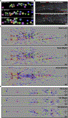
(A) The emission for five distinguishable fluorophores. Each fluorophore’s excitation wavelength is listed in parentheses. (B) Fluorophores are converted into pseudo colors to construct a primary color palette. Three fluorophores are designated as landmarks and pseudo colored to construct an RGB color palette: mNeptune2.5 is pseudo-colored red, CyOFP1 is pseudo-colored green, and mTagBFP2 is pseudo-colored blue. The fluorophore TagRFP-T is used as a panneuronal marker. The fluorophores GFP/CFP/YFP/GCaMP are reserved for reporters of gene expression or neuronal activity. TagRFP-T and GFP/CFP/YFP/GCaMP are visualized separately from the RGB landmarks to avoid confusion. They can be assigned any pseudo color. (C) An example of how to stably pseudo color neurons, across animals. A set of reporters (rows), with stable neuronal expression (columns), are used to drive the fluorophores (table elements). NeuroPAL colors (last row) result from the combined patterns of reporter-fluorophore expression. The panneuronal reporter is expressed in all neurons. The remaining reporters have differential neuronal expression patterns and are used to drive the pseudo-colored landmark fluorophores. Combinations of these differentially expressed reporters assign stable and distinguishable pseudo colors to neurons. For example, reporter 3 drives the red landmark fluorophore (mNeptune2.5), to color neurons 2–4 with distinguishable red intensities and contributes to the blended coloring of several other neurons. In contrast, neuron 1 does not express any of the landmark fluorophores but is still marked by the panneuronal reporter. (D) NeuroPAL scales this concept to 41 reporters that, in combination, disambiguate every neuron in C. elegans and thus generate, a single stereotyped color map across all animals (see Table S1 for data). Seven of the NeuroPAL reporters use a self-cleaving peptide sequence (T2A) to simultaneously drive expression of two different colors. (E) Young adult NeuroPAL worms have a deterministic color map that remains identical across all animals (see Videos S1 and S2 for 3D images). Each neuron is distinguishable from its neighbors via color. All worm ganglia are shown. All images may employ histogram adjustments to improve visibility. Images without a transmitted light channel (e.g., Nomarski) may further be adjusted with a gamma of ~0.5 to improve visibility on the dark background. See also Table S1 and Videos S1 and S2. See Figures S1 and Table S2 for phenotypic analysis.
Figure 2.
Figure 2.. Neuron Locations and Their Positional Variability
(A) Neuron locations and variability, in the retrovesicular ganglion, taken from electron micrographs of three adult hermaphrodites N2S, N2T, and N2U (Hall and Altun, 2007; White et al., 1986). (B) An example of substantial positional variability. The OLL left (OLLL) and right (OLLR) neurons, within a single animal, should share equivalent positions. Instead they show substantial anterior-posterior displacement relative to each other. The transgenic reporters and their pseudo colors are noted on the figure. (C and D) Canonical neuron locations (filled circles displaying the NeuroPAL colors) and their positional variability (encircling ellipses with matching colors) for all ganglia, as determined by NeuroPAL (otIs669, strain OH15262) (see Table S3 for data). Positional variability is shown as the 50% contour for neuronal location (measured as a Gaussian density distribution), sliced within a 2D plane (Methods S1). We show both the left-right and dorsal-ventral planes to provide a 3D estimation of positional variability. (C) Left, right, and ventral views of the head neuron positions. OLLR exhibits over twice the positional variability of OLLL in its anterior-posterior axis, echoing the displacement seen with the non-NeuroPAL transgene in (B). (D) Left, right, and ventral views of the tail neuron positions. See also Table S3 and Methods S1.
Figure 3.
Figure 3.. Expression of All Metabotropic Neurotransmitter Receptors
(A–H) NeuroPAL is used to identify the GFP expression patterns for all metabotropic neurotransmitter receptors (Table S4): the three acetylcholine receptors (A) GAR-1, (B) GAR-2, and (C) GAR-3; the two GABA receptors (D) GBB-1 and (E) GBB-2; and the three glutamate receptors (F) MGL-1, (G) MGL-2, and (H) MGL-3. See also Table S4.
Figure 4.
Figure 4.. Metabotropic Neurotransmitter Communication Network
(A) Expression of the metabotropic neurotransmitter receptors incorporated into the existing, anatomically defined connectome. Rows are presynaptic neurons (organized by neurotransmitter). Columns are postsynaptic neurons (organized by neuron type). Cognate synaptic connections (where the presynaptic neurotransmitter matches the postsynaptic metabotropic receptor) are marked by a colored dot (see legend for neurotransmitter coloring). All other synaptic connections are marked by a black dot. Metabotropic autapses with cognate self-connectivity are circled. (B) The complete GABA communication network: all presynaptic GABA neurons and their corresponding postsynaptic ionotropic GABAA and metabotropic GABAB expressing neurons. Synaptic connections are marked by a colored dot to indicate postsynaptic GABAA and/or GABAB receptors (see legend for receptor-type coloring). GABA autapses are circled. All GABAergic autapses express GABAB receptors and none express GABAA receptors. (C) Metabotropic (and ionotropic GABAA) receptor expression as a percentage of all neurons. Metabotropic receptors are expressed in almost all neurons. (D) Synaptic connections with cognate neurotransmitter receptors as a percentage of the total synapses associated with each neurotransmitter. Metabotropic communication is extensive and these percentages are robust when removing weak synaptic connections by thresholding for at least 1, 5, or 10 synapses. A polyadic synapse that connects AVF (a neuron with no means of releasing its GABA) onto AIM and several cognate GABAB expressing neurons, reduces our count of GABA synaptic signaling to just under 100%. (E) Neurons expressing a neurotransmitter receptor that have no presynaptic partners expressing their cognate neurotransmitter. The absence of presynaptic GABAergic partners for over 31% of the GABAA and 37% of the GABAB receptor-expressing neurons suggests substantial extrasynaptic GABA signaling. For each neurotransmitter, we show the percentage of neurons lacking a cognate presynaptic partner relative to all neurons expressing the neurotransmitter receptor. Removing weak synaptic connections increases the potential for extrasynaptic signaling. (F) The types of neurons expressing a neurotransmitter receptor that have no presynaptic partners expressing their cognate neurotransmitter (suggesting extrasynaptic signaling). We show the neuron type percentages. Sensory neurons represent the majority and are robust against removing weak synaptic connections. Neurons are often categorized as multiple types and thus the sum of percentages can exceed 100%. (G) Ionotropic versus metabotropic GABA communication. Over 60% of GABA connections share both GABAA and GABAB receptors at their postsynaptic sites. The remaining nearly 40% of connections are accounted for solely by metabotropic GABAB at the postsynaptic sites. These percentages remain robust when removing weak synaptic connections. Metabotropic receptor abbreviations: acetylcholine, mAch; glutamate, mGlu; GABA, GABAB; all, mTotal. The ionotropic GABA receptor is abbreviated GABAA. See Table S4 for data.
Figure 5.
Figure 5.. Mutant Analysis of the Conserved Transcription Factors PAG-3/Gli and EOR-1/PLZF
(A and B) Alterations in NeuroPAL coloring reveal neurons with altered fate in pag-3(−) mutant backgrounds: the AVE pre-motor interneuron gains blue-color expression, the PVR interneuron loses all green-color expression, and the VA and VB motor neurons are missing entirely. Statistical bar graph colors in (B) are sampled from the images in (A). (C) PAG-3/Gli expression as assessed with a fosmid reporter (wgIs154): ADA*, ALM, AQR*, AVD*, AVE*, AVF, AVH*, AVJ*, AVM, BDU, DVC*, I1*, I2*, I6*, PLM, PQR*, PVC*, PVM, PVQ*, PVR*, PVW*, RID*, RIG, RMG*, VA11–12, and URY* (*previously unpublished expression). (D) EOR-1/PLZF has broad, likely ubiquitous, expression, as assessed with a fosmid reporter (wgIs81). (E–G) The NeuroPAL blue reporters in RMED/V are ggr-3, pdfr-1, and unc-25. (E and F) NeuroPAL exhibits total loss of its RMED/V blue-color expression in eor-1(−) mutant backgrounds but retains panneuronal marker expression (shown in white), indicating the preservation of RMED/V neuronal fate. (G) RMED/V fate analysis in eor-1(−) mutants with unc-25 (otIs514) and unc-47 (otIs565) reporter transgenes. See Table S5 for data.
Figure 6.
Figure 6.. Whole-Brain Neuronal Activity Imaging of Taste and Odor Responses
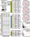
(A) C. elegans were subjected to three chemosensory stimuli: a repulsive taste (160 mM NaCl) and two attractive odors (10−4 2-butanone and 10−4 2,3-pentanedione). (B) Animals were immobilized inside a microfluidic chip. Stimuli were delivered in chemotaxis buffer. Each animal was imaged using a spinning disk confocal microscope with four excitation lasers. The NeuroPAL color map was imaged to identify all neurons. Thereafter, brainwide activity was recorded via the panneuronal calcium sensor GCaMP6s (Videos S3 and S4). (C) Peak neuronal activity, before and during stimulus presentation, for 109 head neuron classes and subclasses. (D–F) Neuronal activity traces for selected (D) sensory neurons, (E) interneurons, and (F) pharyngeal neurons that responded to stimuli. The 10-s stimulus delivery period is indicated by the vertical colored bar. Black activity traces represent all neurons combined into one representative group. Colored activity traces divide neurons into groups exhibiting known asymmetric responses: stereotyped asymmetries between the ASEL and ASER neuron pair and stochastic asymmetries between the AWCON and AWCOFF neuron pair. Significant responses (p or q ≤ 0.05) are highlighted by bold borders. “Post,” significant post-stimulus response; ns, no significant response. (F–I) Average pairwise correlations between 189 neurons in the 30 s following onset of (G) NaCl, (H) 2-butanone, and (I) 2,3-pentanedione. All three correlation maps are presented on the same axes, determined by clustering the full-time-course correlations. The set of correlated and anti-correlated neurons differs for each stimulus presentation. (J and K) Comparison of functional activity to the connectome. We observe minimal correspondence between synapse counts and pairwise-functional-activity correlations for the (J) head and (K) tail. See Table S6 for primary data. See also Figures S1 and S2.
Figure 7.
Figure 7.. NeuroPAL Software: An Algorithm for Semi-automated Neuronal Identification and an Algorithm to Generate Optimal-Coloring Solutions for Cell Identification
(A–C) The algorithm used for semi-automated neuronal identification. (A) Raw images are filtered to remove non-neuronal fluorescence and neurons are detected in the filtered image. Detected neurons are identified by matching them to a statistical atlas of neuronal colors and positions (Table S3). (B and C) Semi-automated neuronal identification accuracy begins at 86% for the head and 94% for the tail. Manually identifying eight neurons raises the head accuracy above 90%. Overall accuracy is displayed as a black line. Accuracy for each ganglion is displayed as a dotted, colored line (see legend). Many of the neurons and ganglia have high identification accuracy and confidence. The ventral ganglion is a problematic area, likely due to the density and high positional variance therein. (D and E) The algorithm used to generate optimal-coloring solutions for cell identification—for any collection of cells in any organism. We show simulations of two approximately optimal alternatives to NeuroPAL, one that permits as many reporters as NeuroPAL (D) and one that restricts the transgene to only 3 reporters (E). With the exception of the number of reporters, both alternatives were generated using parameters similar to NeuroPAL: three landmark fluorophores, where each fluorophore is distinguishable at three intensities (high, medium, and low). Reporters were chosen by the algorithm from those available in WormBase, a community-curated database of cell-specific reporter expression. Similar databases are available for other model organisms (e.g., fly, fish, and mouse). We evaluated the two NeuroPAL alternatives by computing the percentage of their color violations, defined as neighboring neuron pairs with indistinguishable colors. See Methods S1 and S2 for algorithmic details and validation. See also Figure S3 and Table S3

References

    1. Ahrens MB, and Engert F (2015). Large-scale imaging in small brains. Curr. Opin. Neurobiol. 32, 78–86. - PMC - PubMed
    1. Ahrens MB, Orger MB, Robson DN, Li JM, and Keller PJ (2013). Whole-brain functional imaging at cellular resolution using light-sheet microscopy. Nat. Methods 10, 413–420. - PubMed
    1. Bamber BA, Beg AA, Twyman RE, and Jorgensen EM (1999). The Caenorhabditis elegans unc-49 locus encodes multiple subunits of a heteromultimeric GABA receptor. J. Neurosci. 19, 5348–5359. - PMC - PubMed
    1. Bargmann CI, and Horvitz HR (1991). Chemosensory neurons with overlapping functions direct chemotaxis to multiple chemicals in C. elegans. Neuron 7, 729–742. - PubMed
    1. Bargmann CI, and Marder E (2013). From the connectome to brain function. Nat. Methods 10, 483–490. - PubMed

Publication types

MeSH terms

Substances