. 2020 Dec 29;33(13):108553.
doi: 10.1016/j.celrep.2020.108553.

Airway-Associated Macrophages in Homeostasis and Repair

Affiliations

Airway-Associated Macrophages in Homeostasis and Repair

Anna E Engler et al. Cell Rep. .

Abstract

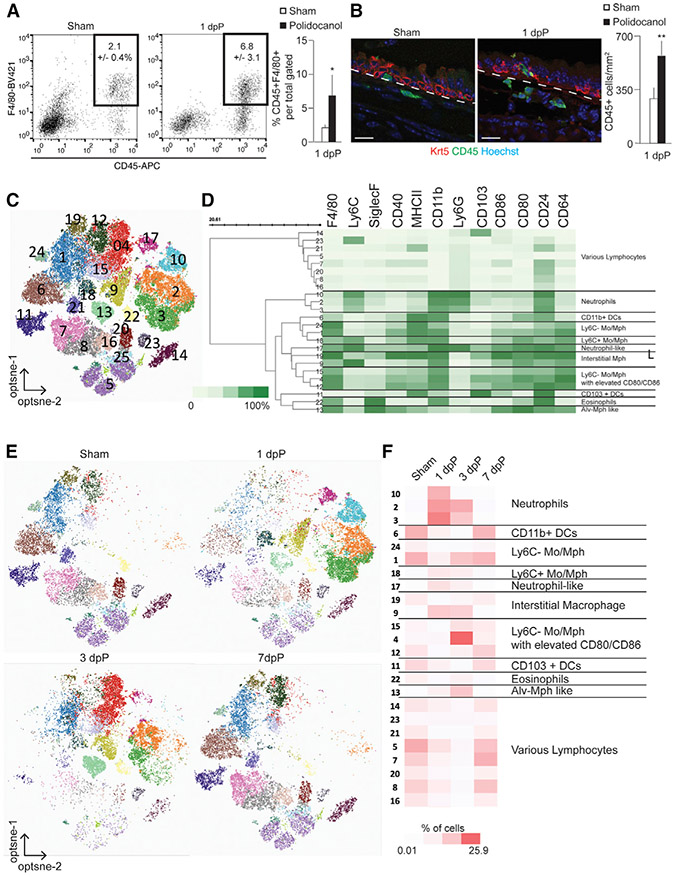
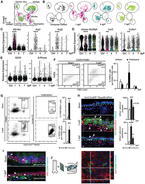
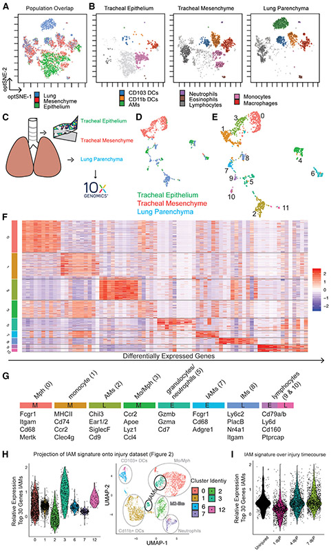
There is an increasing appreciation for the heterogeneity of myeloid lineages in the lung, but relatively little is known about populations specifically associated with the conducting airways. We use single-cell RNA sequencing, flow cytometry, and immunofluorescence to characterize myeloid cells of the mouse trachea during homeostasis and epithelial injury/repair. We identify submucosal macrophages, similar to lung interstitial macrophages, and intraepithelial macrophages. Following injury, there are early increases in neutrophils and submucosal macrophages, including M2-like macrophages. Intraepithelial macrophages are lost after injury and later restored by CCR2+ monocytes. We show that repair of the tracheal epithelium is impaired in Ccr2-deficient mice. Mast cells and group 2 innate lymphoid cells are sources of interleukin-13 (IL-13) that polarize macrophages and directly influence basal cell behaviors. Their proximity to the airway epithelium establishes these myeloid populations as potential therapeutic targets for airway disease.

Keywords: airway; macrophages; niche; regeneration; trachea.


Conflict of interest statement

Declaration of Interests S.A.M. received sponsored research funding from Janssen Pharmaceuticals.

Figures

Figure 1.
Figure 1.. Characterization of Tracheal Macrophages
(A) FACS analysis of CD45+;F4/80+ myeloid cells after 1 dpP; (n = 4). (B) Immunofluorescence and quantifications of CD45+ cells (green) near basal cells (KRT5, red) following polidocanol-induced injury. Scale bar, 25 μm. (C) Unsupervised clustering of all cells combined from sham and from 1, 3, and 7 dpP based on 15-marker myeloid panel; (n = 1/time point, consistent of 2 animals). (D) Heatmap of marker expression data, scaled from 0% to 100% of normalized, arcsin value of fluorescent intensity. (E) Cells isolated on indicated days post-injury, visualized by cluster assignment. (F) Relative abundance of cells from 24 distinct clusters across injury and repair, scaled 0.01% to 25.9% of total cells in analysis. See also Figure S1 and Table S1.
Figure 2.
Figure 2.. Dynamic Features of Tracheal Myeloid Populations after Injury
(A) UMAP visualization of all SCRNA-seq data across airway injury and repair overlaid (3 animals pooled into 1 sample for each); captured cells: ctrl = 458, 1 dpP = 1,625, 4 dpP = 1,507, 7 dpP = 1,717. (B) UMAP visualization of SCRNA-seq data on indicated days post-injury. (C) Violin plots of M2-like macrophage gene signature, Arg1 and Arg2. (D) Violin plots of classic monocyte/macrophage gene signature, Ccr2 and Cx3cr1. (E) Violin plots of cell cycle genes associated with G2/M and S phase. (F) Representative FACS plot and quantification of YARG expression gated on CD45+;F4/80+ (n = 3 mice per time point). (G) FACS analysis and quantification of Ccr2-CreERT2;ROSA-tdTom+ cells from YARG animals after tamoxifen administration and sham or polidocanol treatment (n = 3, mice per time point). (H) Immunofluorescence analysis of Ccr2-CreERT2;ROSA-tdTom animals; Krt5 (basal cells, green), Ccr2-CreERT2;ROSA-tdTom lineage trace (red), and Höechst nuclear stain (blue); scale bar, 15 μm. (I) Histology of wild-type animals; Krt5 (red), CD45 (green), nuclear stain Höechst, scale bar, 15 μm. (J) Confocal image of peeled epithelial sheet showing F4/80+ myeloid cells (green) and Krt5+ epithelial basal cells (red), Höechst nuclear stain. See also Figure S2 and Tables S2 and S4.
Figure 3.
Figure 3.. Myeloid Cells Isolated from the Epithelium, Mesenchyme, and Lung Parenchyma Have Distinct Transcriptional and Translational Identities
(A) Unsupervised clustering of 15-marker myeloid FACS panel, overlay (n = 5/tissue pooled). (B) Visualization of cells isolated from epithelium (left), mesenchyme (middle), and lung (right) after pooled unsupervised clustering. (C) Schematic representation of samples isolated for SCRNA-seq (n = 5/tissue animals pooled). (D) UMAP colored for tissue of origin. (E) Unsupervised Louvaine clustering at resolution 0.25 of SCRNA-seq data. (F) Heatmap of differentially expressed genes in myeloid clusters. (G) Cluster assignment based on selected transcripts and annotation of tissue of origin (E, epithelium; M, mesenchyme; L, lung). (H) Projection of IAM gene signature (cluster 7; Figure 3F) onto single-cell injury dataset (Figures 2A-2E). (I) Projection of IAM gene signature onto injury time points. See also Figure S3 and Tables S1, S3, and S4.
Figure 4.
Figure 4.. CCR2+ Myeloid Cells Promote Basal-Cell-Mediated Epithelial Repair
(A) Cocultures (n = 4) of basal cells and CCR2+ cells or Arg1+ cells isolated from sham or polidocanol conditions. (B) FACS analysis of IL-4Rα expression on basal cells (n = 3). (C) Schematic representation of basal cell isolation, infection, and manipulation. (D and E) Quantification (D) and bright field (E) of culture of IL-4Rα+/+ (n = 3) and IL-4Rα−/− (n = 3) basal cells in the presence of IL-4 or IL-13 and quantification of sphere size after 10 days of culture. (F) Epithelial repair in CCR2-deficient mice and heterozygous controls, 3 dpP and 7 dpP. Heterozygote, sham: n = 2, injury: n = 3; homozygote sham: n = 4, injury: n = 3. Scale bars, 25 μm. Quantification of acetylated α-tubulin+ ciliated cells per mm2 as a metric of repair. See also Figure S4.

References

    1. Anderson KG, Mayer-Barber K, Sung H, Beura L, James BR, Taylor JJ, Qunaj L, Griffith TS, Vezys V, Barber DL, and Masopust D (2014). Intravascular staining for discrimination of vascular and tissue leukocytes. Nat. Protoc 9, 209–222. - PMC - PubMed
    1. Belkina AC, Ciccolella CO, Anno R, Halpert R, Spidlen J, and Snyder-Cappione JE (2019). Automated optimized parameters for T-distributed stochastic neighbor embedding improve visualization and analysis of large datasets. Nat. Commun 10, 5415. - PMC - PubMed
    1. Borthwick DW, Shahbazian M, Krantz QT, Dorin JR, and Randell SH (2001). Evidence for stem-cell niches in the tracheal epithelium. Am. J. Respir. Cell Mol. Biol 24, 662–670. - PubMed
    1. Byrne AJ, Mathie SA, Gregory LG, and Lloyd CM (2015). Pulmonary macrophages: key players in the innate defence of the airways. Thorax 70, 1189–1196. - PubMed
    1. Chakarov S, Lim HY, Tan L, Lim SY, See P, Lum J, Zhang X-M, Foo S, Nakamizo S, Duan K, et al. (2019). Two distinct interstitial macrophage populations coexist across tissues in specific subtissular niches. Science 363, 80. - PubMed

Publication types

MeSH terms

LinkOut - more resources