. 2020 Dec 29;33(13):108569.
doi: 10.1016/j.celrep.2020.108569.

DNA and RNA Cleavage Complexes and Repair Pathway for TOP3B RNA- and DNA-Protein Crosslinks

Affiliations

DNA and RNA Cleavage Complexes and Repair Pathway for TOP3B RNA- and DNA-Protein Crosslinks

Sourav Saha et al. Cell Rep. .

Abstract

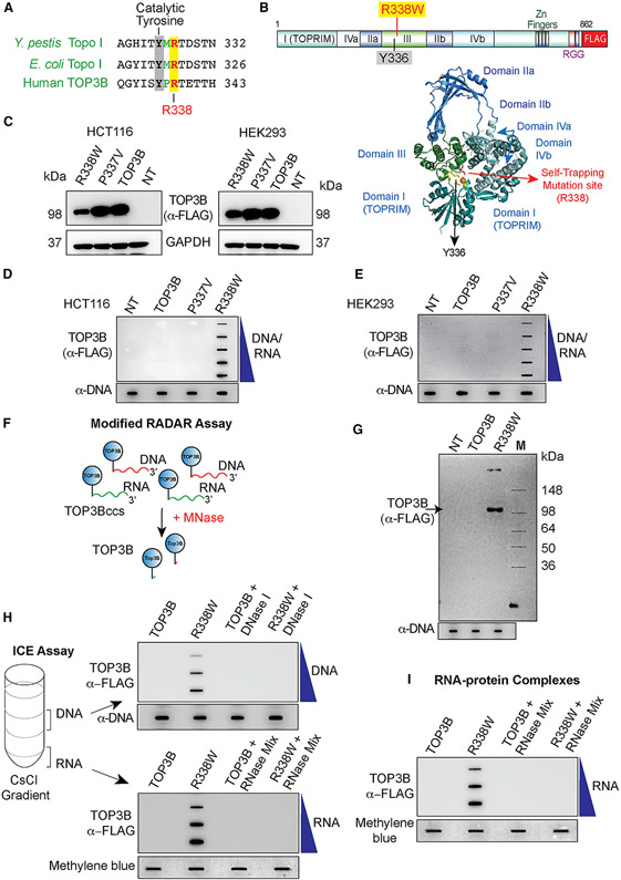
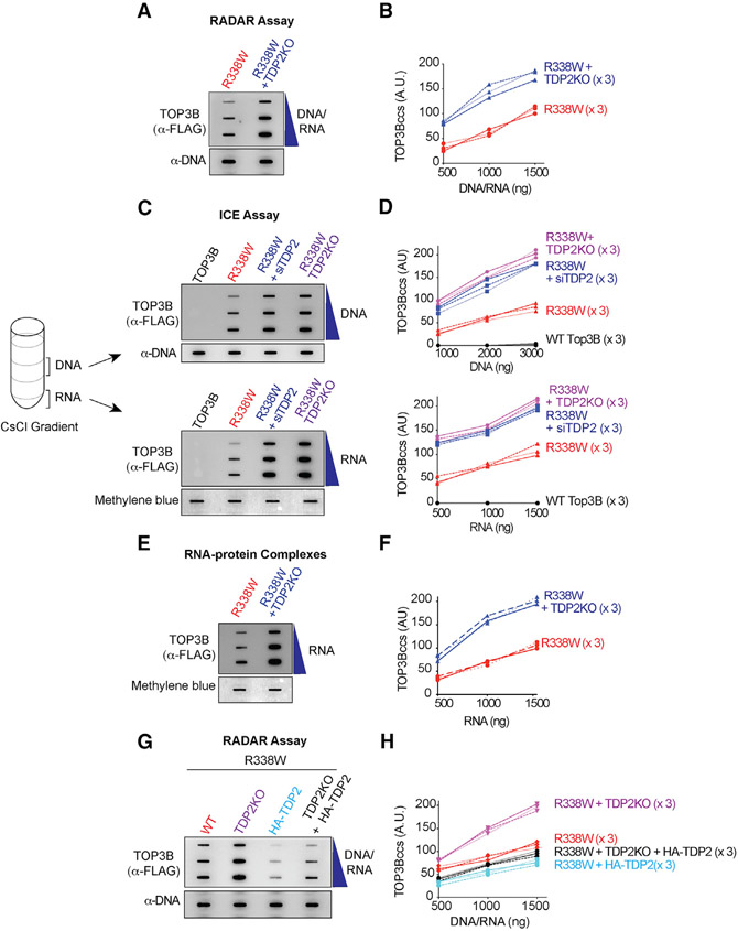
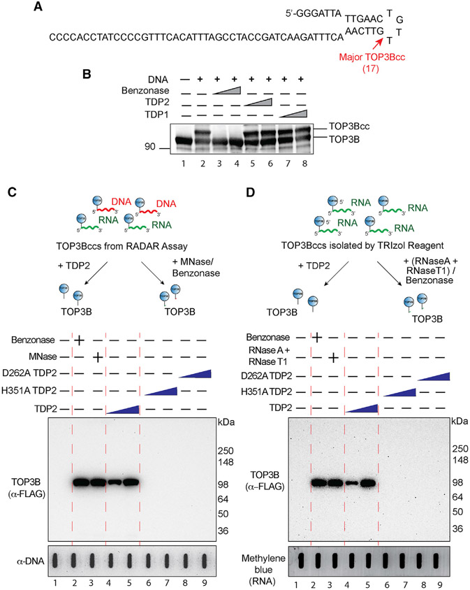
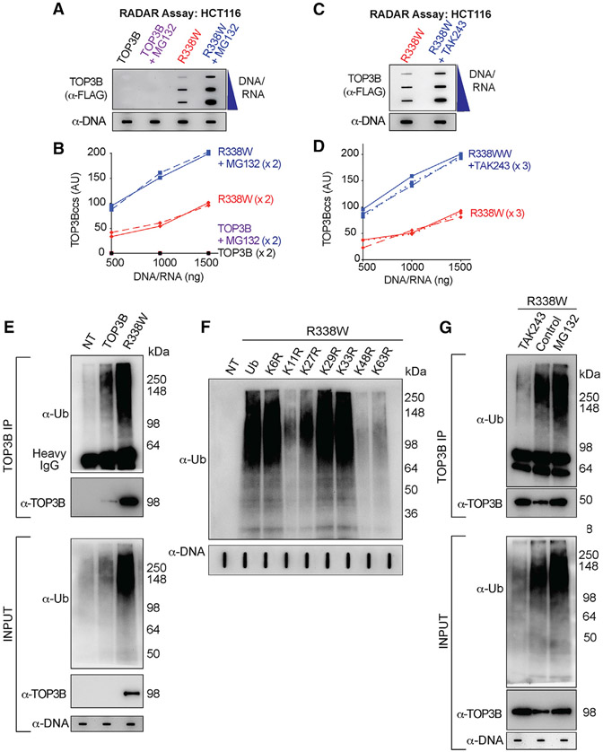
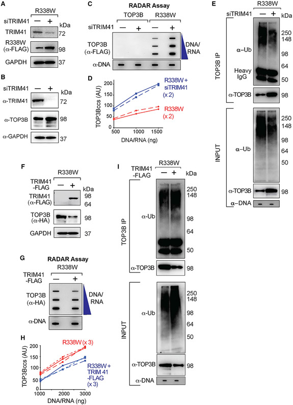
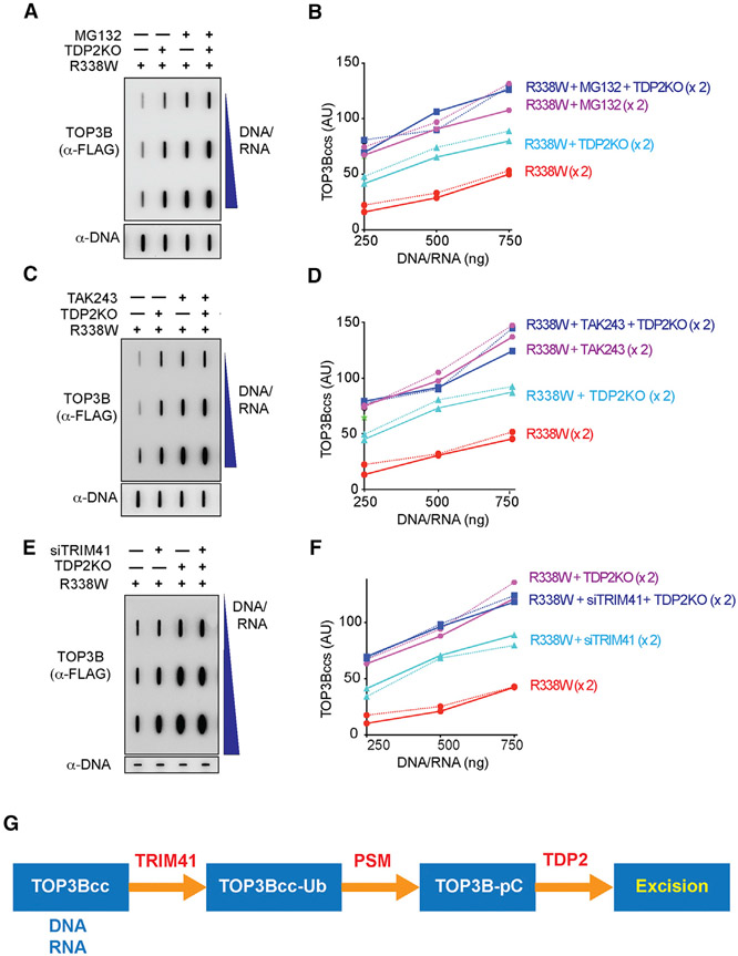
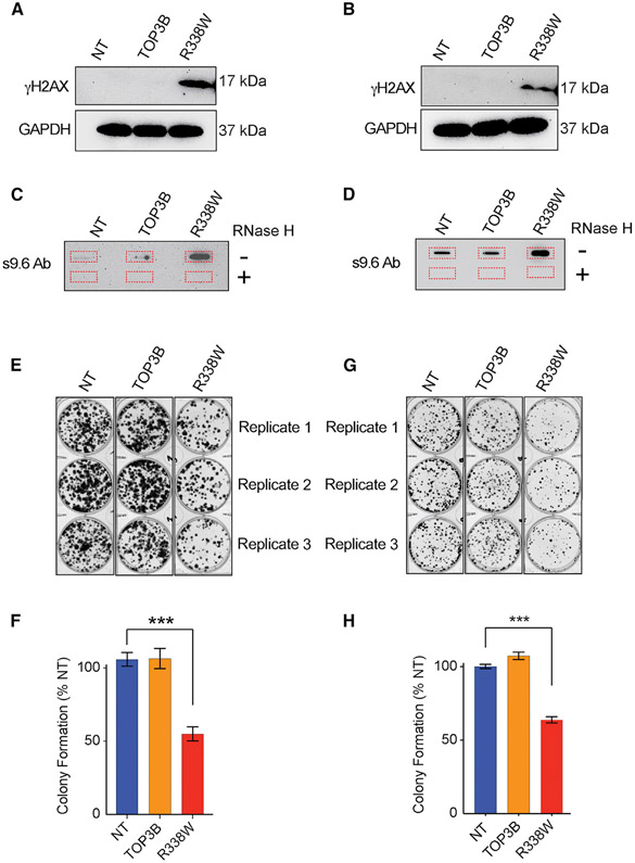
The present study demonstrates that topoisomerase 3B (TOP3B) forms both RNA and DNA cleavage complexes (TOP3Bccs) in vivo and reveals a pathway for repairing TOP3Bccs. For inducing and detecting cellular TOP3Bccs, we engineer a "self-trapping" mutant of TOP3B (R338W-TOP3B). Transfection with R338W-TOP3B induces R-loops, genomic damage, and growth defect, which highlights the importance of TOP3Bcc repair mechanisms. To determine how cells repair TOP3Bccs, we deplete tyrosyl-DNA phosphodiesterases (TDP1 and TDP2). TDP2-deficient cells show elevated TOP3Bccs both in DNA and RNA. Conversely, overexpression of TDP2 lowers cellular TOP3Bccs. Using recombinant human TDP2, we demonstrate that TDP2 can process both denatured and proteolyzed TOP3Bccs. We also show that cellular TOP3Bccs are ubiquitinated by the E3 ligase TRIM41 before undergoing proteasomal processing and excision by TDP2.

Keywords: DNA repair; TDP2; TRIM41; topoisomerase; ubiquitin-proteasome.


Conflict of interest statement

Declaration of Interests The authors declare no competing interests.

Figures

Figure 1.
Figure 1.. TOP3B Forms TOP3Bccs Both with DNA and RNA in Cells Transfected with R338W-TOP3B
(A) Alignment of the active site regions of Y. pestis Topo I, E. coli Topo I, and human TOP3B. (B) Structure of human TOP3B and ribbon representation of human TOP3B (amino acid [aa] residues 1–612) (Goto-Ito et al., 2017) with the active site Y336 and the self-trapping mutation site (R338). (C) Ectopic expression of wild-type (WT) TOP3B, P337V, and R338W-TOP3B following transfection of HCT116 and HEK293 cells with the indicated TOP3B constructs for 72 h. Western blotting with anti-FLAG antibody. (D and E) Detection of TOP3Bccs by RADAR assay in cells transfected with the indicated plasmid constructs for 72 h. TOP3Bccs were detected with anti-FLAG antibody. Equal loading was determined by slot blotting and probing with anti-dsDNA antibody. The figure is representative of three independent experiments. NT, mock-transfected cells. (F) FLAG-tagged TOP3B (blue circles) cellular TOP3Bccs in DNA (red) and RNA (green) were digested with micrococcal nuclease (MNase) followed by SDS-PAGE and immunoblotting with anti-FLAG antibody. (G) Modified RADAR assay in HEK293 cells transfected with WT or R338W-TOP3B for 72 h. TOP3B was detected with anti-FLAG antibody. Equal loading was tested by slot blotting and probing with anti-dsDNA antibody. (H) TOP3Bccs in DNA and RNA of HEK293 cells transfected for 72 h with WT or R338W-TOP3B. Cesium chloride gradient ultracentrifugation was performed to separate DNA and RNA (middle and bottom of the gradient, respectively). DNA and RNA fractions were treated with excess RNase A (200 μg/mL) and RNase T1 (200 Units/ml) or DNase I (10 units) as indicated. DNA and RNA fractions were slot blotted and TOP3Bccs detected with anti-FLAG antibody. Equal loading was determined by slot blotting and probing with anti-dsDNA antibody (DNA) or methylene blue staining (RNA). The figure is representative of three independent experiments. (I) TOP3Bccs in RNA of HEK293 cells transfected with WT or R338W-TOP3B for 72 h. Covalent protein-RNA adducts were isolated using TRIzol (Thermo Scientific) and treated with excess RNase A (200 μg/mL) and RNase T1 (200 units/ml) as indicated. Slot-blotted TOP3Bccs were detected with anti-FLAG antibody. Equal loading was determined by slot blotting and methylene blue staining. The figure is representative of three independent experiments. See also Figure S1.
Figure 2.
Figure 2.. TDP2 Excises Cellular TOP3Bccs from Both DNA and RNA
(A) WT or TDP2KO HCT116 cells were transfected with R338W-TOP3B. After 72 h, protein-nucleic acid adducts were isolated by RADAR assay. Slot-blotted TOP3Bccs were detected with anti-FLAG antibody. Loading was tested by slot blotting and probing with anti-dsDNA antibody. (B) Quantitation of TOP3Bccs from three independent experiments as shown in (A). TOP3Bccs were measured by densitometric analyses of slot-blot signals and plotted as a function of total nucleic acid (DNA and RNA) concentration. (C) HCT116 WT and TDP2KO cells were transfected with WT-TOP3B or R338W-TOP3B and siTDP2 constructs as indicated. After 72 h, ICE bioassays were performed to isolate DNA and RNA fractions. TOP3Bccs were detected with anti-FLAG antibody. Loading was tested with anti-dsDNA antibody or methylene blue staining (RNA). (D) Quantitation of TOP3Bcc in three independent experiments as shown in (C). (E) WT and TDP2KO HCT116 cells were transfected with FLAG-tagged R338W-TOP3B. After 72 h, protein-RNA adducts were isolated using TRIzol. TOP3Bccs were detected with anti-FLAG antibody. Loading was tested by methylene blue staining. (F) Quantitation of TOP3Bcc in RNA in three independent experiments as shown in (E). (G) Ectopic expression of TDP2 reduces TOP3Bccs. WT and TDP2KO HCT116 cells were transfected with FLAG-tagged R338W-TOP3B alone or co-transfected with HA-tagged TDP2. After 72 h, TOP3Bccs were detected with anti-FLAG antibody. Loading was tested with anti-dsDNA antibody. (H) Quantitation of TOP3Bccs in three independent experiments as shown in (G). See also Figure S2.
Figure 3.
Figure 3.. Recombinant Human TDP2 Excises Denatured but Not Native TOP3Bcc
(A) Oligonucleotide substrate (69-mer) with main TOP3B site (17 nucleotides from the 5′ end). (B) Recombinant human TDP2 does not excise native TOP3Bccs. The oligonucleotide (300 nM) was incubated with recombinant human TOP3B (4 μM). TOP3Bcc formation results in a slower migrating band (lane 2) (for full gel and additional minor bands see Figure S3A). TOP3Bccs were incubated with 1 or 3 μM recombinant TDP1 (lanes 7 and 8) or TDP2 (lanes 5 and 6). Benzonase (3 and 9 units, lanes 3 and 4,) was used as a positive control to degrade the oligonucleotide and release TOP3B. Samples were immunoblotted with anti-TOP3B antibody after SDS-PAGE. (C) Recombinant human TDP2 excises denatured cellular DNA and RNA TOP3Bccs. HEK293 cells were transfected with FLAG-tagged R338W-TOP3B, and protein-nucleic acid adducts were recovered by RADAR assay. After incubation with recombinant TDP2 (3 and 6 μM, lanes 4 and 5), H351A TDP2 (3 and 6 μM, lanes 6 and 7), D262A TDP2 (3 and 6 μM, lanes 8 and 9), benzonase (250 units, lane 2), or MNase (300 units, lane 3), released TOP3B was detected by immunoblotting with anti-FLAG antibody after SDS-PAGE. Loading was tested by slot blotting and probing with anti-dsDNA antibody. (D) Recombinant human TDP2 excises cellular RNA TOP3Bccs. HEK293 cells were transfected with R338W-TOP3B. Covalent protein-RNA adducts were isolated using TRIzol. After incubation with recombinant TDP2 (3 and 6 μM, lanes 4 and 5), H351A TDP2 (3 and 6 μM, lanes 6 and 7), D262A TDP2 (3 and 6 μM, lanes 8 and 9), benzonase (250 units, lane 2), or an excess amount of RNase A (200 μg/mL) and RNase T1 (200 units/ml) mix (lane 3), released TOP3B was detected by immunoblotting with anti-FLAG antibody after SDS-PAGE. Loading was tested by methylene blue staining (RNA). See also Figure S3.
Figure 4.
Figure 4.. Cellular TOP3Bccs Are Ubiquitinated and Degraded by the Proteasomal Pathway
(A) Proteasome inhibition enhances cellular TOP3Bccs. HCT116 cells were transfected with WT TOP3B and R338W-TOP3B for 72 h. Before harvest, cells were treated with MG132 (10 μM, 2 h). TOP3Bccs were detected by using anti-FLAG antibody. Loading was tested with anti-dsDNA antibody. The figure is representative of two independent experiments. (B) Quantitation of TOP3Bcc in two independent experiments as shown in (A). (C) Ubiquitination inhibition enhances cellular TOP3Bccs. HCT116 cells transfected with FLAG-tagged R338W-TOP3B for 72 h were treated with the UAE inhibitor TAK243 (10 μM, 2 h) before harvest. TOP3Bccs were detected with anti-FLAG antibody. Loading was tested by slot blotting and probing with anti-dsDNA antibody. The figure is representative of three independent experiments. (D) Quantitation of TOP3Bcc in three independent experiments as shown in (C). (E) Ubiquitination of cellular TOP3Bccs. RADAR assay samples were prepared from NT HCT116 cells or cells transfected with FLAG-tagged WT-TOP3B or R338W-TOP3B. Equal amounts of RADAR assay samples were immunoprecipitated (IPed) with anti-TOP3B antibody. IPed samples and RADAR samples were digested with MNase, resolved on SDS-PAGE, and immunoblotted with anti-Ub and anti-TOP3B antibodies. Loading (input) was tested with anti-dsDNA antibody. (F) TOP3Bcc ubiquitination involves proteasomal-specific linkages to lysines K11, K27, K48, and K63. HCT116 cells were co-transfected with R338W-TOP3B and HA-tagged WT or mutant ubiquitin constructs for 72 h. RADAR samples were treated with MNase and probed with anti-Ub antibody. Loading was tested with anti-dsDNA antibody. (G) Inhibition of TOP3Bcc ubiquitination by the UAE inhibitor TAK243 and enhancement by the proteasome inhibitor MG132. HCT116 cells transfected with FLAG-tagged R338W-TOP3B for 72 h were treated with either MG132 (10 μM, 2 h) or TAK243 (10 μM, 2 h), RADAR assay samples were isolated and IPed with an anti-TOP3B antibody. IP samples and the input RADAR assay samples were digested with MNase, resolved on SDS-PAGE and immunoblotted with anti-Ub and anti-TOP3B antibodies. Equal loading was tested by slot blotting and probing with anti-dsDNA antibody. See also Figure S4.
Figure 5.
Figure 5.. TRIM41 Acts as a Ubiquitin Ligase for TOP3Bccs and Promotes the Repair of TOP3Bccs
(A) Immunoblots showing TRIM41 and R338W-TOP3B expression after TRIM41 downregulation (GAPDH as loading control). HCT116 cells were either transfected with a FLAG-tagged R338W-TOP3B plasmid construct alone or co-transfected with a siTRIM41construct for 72 h. (B) Immunoblots showing effect of TRIM41 depletion on endogenous TOP3B level. GAPDH was included as loading control. HCT116 cells were transfected with siTRIM41construct for 72 h. (C) HCT116 cells were transfected with FLAG-tagged WT- or R338W-TOP3Bs or co-transfected with siTRIM41construct for 72 h. TOP3Bcc were detected with anti-FLAG antibody. Equal loading was tested with anti-dsDNA antibody. The figure is representative of two independent experiments. (D) Quantitation of TOP3Bccs in two independent RADAR assays as shown in (C). (E) HCT116 cells were transfected with FLAG-tagged R338W-TOP3B alone or with siTRIM41 construct. After 72 h, equal amounts of RADAR assay samples were IPed with anti-TOP3B antibody. IP samples and the input RADAR assay samples were digested with MNase, resolved on SDS-PAGE and immunoblotted with anti-Ub and anti-TOP3B antibodies. Loading of input RADAR samples was tested with anti-dsDNA antibody. (F) Immunoblots showing TRIM41 and R338W-TOP3B expression after transfection of HCT116 cells with HA-tagged R338W-TOP3B plasmid construct alone or with FLAG-tagged TRIM41. GAPDH served as loading control. (G) Reduced TOP3Bccs upon TRIM41 overexpression. HCT116 cells were transfected with HA-tagged R338W-TOP3B alone or co-transfected with FLAG-tagged TRIM41 for 48 h. TOP3Bccs were detected with anti-HA antibody. Loading was tested by with anti-dsDNA antibody. The figure is representative of three independent experiments. (H) Quantitation of TOP3Bcc formation in three independent RADAR assays as shown in (F). (I) Increased TOP3Bcc ubiquitination upon transfection with TRIM41. HCT116 cells were either transfected with HA-tagged R338W-TOP3B alone or co-transfected with FLAG-tagged TRIM41. After 48 h, RADAR assay samples were IPed with anti-TOP3B antibody. IP samples and the input RADAR assay samples were digested with MNase, resolved on SDS-PAGE, and immunoblotted with anti-Ub and anti-TOP3B antibodies. Loading was tested with anti-dsDNA antibody. See also Figure S5.
Figure 6.
Figure 6.. TDP2-Mediated Repair of TOP3Bccs Is Dependent on Ubiquitination and Proteasomal Processing
(A) WT and TDP2KO HCT116 cells were transfected with FLAG-tagged R338W-TOP3B alone and incubated for 72 h. Before harvest, cells were treated with MG132 (10 μM, 2 h) as indicated. Nucleic acids and protein-nucleic acid adducts were recovered by RADAR assay. TOP3Bccs were detected using anti-FLAG antibody. Equal loading was with anti-dsDNA antibody. The figure is representative of two independent experiments. (B) Quantitation of TOP3Bccs in two independent experiments as shown in (A). (C) WT and TDP2KO HCT116 cells were transfected with FLAG-tagged R338W-TOP3B alone and incubated for 72 h. Before harvest, cells were treated with TAK243 (10 μM, 2 h). Nucleic acids and protein-nucleic acid adducts were recovered by RADAR assay, and TOP3Bccs were detected using anti-FLAG antibody. Equal loading was tested with anti-dsDNA antibody. The figure is representative of two independent experiments. (D) Quantitation of TOP3Bccs in two independent RADAR assays as shown in (C). (E) WT and TDP2KO HCT116 cells were transfected with FLAG-tagged R338W-TOP3B alone or co-transfected with siTRIM41 constructs and incubated for 72 h. TOP3Bccs were detected after RADAR assay with anti-FLAG antibody. Equal loading was tested with anti-dsDNA antibody. The figure is representative of two independent experiments. (F) Quantitation of TOP3Bccs in two independent RADAR assays as shown in (E). (G) Model for the processing of TOP3Bccs by TRIM14, the proteasome (PSM), and TDP2. See also Figure S6.
Figure 7.
Figure 7.. Cellular Consequences of TOP3B Trapping and Identification of RNAs Associated with TOP3B
(A) Trapping of TOP3B results in γH2AX induction. Immunoblots showing γH2AX levels in HEK293 cells transfected with R338W-TOP3B, WT-TOP3B, or NT. (B) Trapping of TOP3B results in γH2AX induction in HCT116 cells transfected with R338W-TOP3B, WT-TOP3B, or mock-transfection reagent (NT). (C) Trapping of TOP3B results in R-loop accumulation. Genomic DNA isolated from HEK293 cells transfected with R338W-TOP3B, WT-TOP3B, or NT was slot blotted and probed with S9.6 antibody. (D) Trapping of TOP3B results in R-loop accumulation in HCT116 cells transfected with R338W-TOP3B, WT-TOP3B, or NT. (E) Representative images of colony formation assay in HEK293 cells after transfection with R338W-TOP3B, WT-TOP3B or NT. (F) Quantitative representation of colony formation assays as shown in (E). Data are provided as means ± standard deviations (SD) (n = 3). ***p ≤ 0.0005 (two-tailed unpaired t test). (G) Representative images of colony formation assay in HCT116 cells after transfection with R338W-TOP3B, WT-TOP3B, or NT. (H) Quantitative representation of colony formation assays as shown in (G). Data are provided as means ± standard deviations (SD) (n = 3). ***p ≤ 0.0005 (two-tailed unpaired t test).

References

    1. Ahmad M, Xue Y, Lee SK, Martindale JL, Shen W, Li W, Zou S, Ciaramella M, Debat H, Nadal M, et al. (2016). RNA topoisomerase is prevalent in all domains of life and associates with polyribosomes in animals. Nucleic Acids Res. 44, 6335–6349. - PMC - PubMed
    1. Ahmad M, Shen W, Li W, Xue Y, Zou S, Xu D, and Wang W (2017a). Topoisomerase 3β is the major topoisomerase for mRNAs and linked to neuro-development and mental dysfunction. Nucleic Acids Res. 45, 2704–2713. - PMC - PubMed
    1. Ahmad M, Xu D, and Wang W (2017b). Type IA topoisomerases can be “magicians” for both DNA and RNA in all domains of life. RNA Biol. 14, 854–864. - PMC - PubMed
    1. Bedford L, Lowe J, Dick LR, Mayer RJ, and Brownell JE (2011). Ubiquitin-like protein conjugation and the ubiquitin-proteasome system as drug targets. Nat. Rev. Drug Discov 10, 29–46. - PMC - PubMed
    1. Castillo-Guzman D, Hartono SR, Sanz LA, and Chédin F (2020). SF3B1-targeted Splicing Inhibition Triggers Global Alterations in Transcriptional Dynamics and R-Loop Metabolism. bioRxiv, 2020.2006.2008.130583.

Publication types

MeSH terms