. 2020 Dec 28;10(1):14.
doi: 10.3390/pathogens10010014.

Genomic Sequencing and Comparison of Sacbrood Viruses from Apis cerana and Apis mellifera in Taiwan

Affiliations

Genomic Sequencing and Comparison of Sacbrood Viruses from Apis cerana and Apis mellifera in Taiwan

Ju-Chun Chang et al. Pathogens. .

Abstract

Sacbrood virus (SBV) was the first identified bee virus and shown to cause serious epizootic infections in the population of Apis cerana in Taiwan in 2015. Herein, the whole genome sequences of SBVs in A. cerana and A. mellifera were decoded and designated AcSBV-TW and AmSBV-TW, respectively. The whole genomes of AcSBV-TW and AmSBV-TW were 8776 and 8885 bp, respectively, and shared 90% identity. Each viral genome encoded a polyprotein, which consisted of 2841 aa in AcSBV-TW and 2859 aa in AmSBV-TW, and these sequences shared 95% identity. Compared to 54 other SBVs, the structural protein and protease regions showed high variation, while the helicase was the most highly conserved region among SBVs. Moreover, a 17-amino-acid deletion was found in viral protein 1 (VP1) region of AcSBV-TW compared to AmSBV-TW. The phylogenetic analysis based on the polyprotein sequences and partial VP1 region indicated that AcSBV-TW was grouped into the SBV clade with the AC-genotype (17-aa deletion) and was closely related to AmSBV-SDLY and CSBV-FZ, while AmSBV-TW was grouped into the AM-genotype clade but branched independently from other AmSBVs, indicating that the divergent genomic characteristics of AmSBV-TW might be a consequence of geographic distance driving evolution, and AcSBV-TW was closely related to CSBV-FZ, which originated from China. This 17-amino-acid deletion could be found in either AcSBV or AmSBV in Taiwan, indicating cross-infection between the two viruses. Our data revealed geographic and host specificities between SBVs. The amino acid difference in the VP1 region might serve as a molecular marker for describing SBV cross-infection.

Keywords: Apis cerana; Apis mellifera; sacbrood disease; sacbrood virus.


Conflict of interest statement

The authors declare there is no conflict of interest involved in this work.

Figures
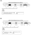
Figure 1
Figure 1
The genomic maps of (A) AmSBV-TW (accession number: MN082651) and (B) AcSBV-TW (accession number: MN082652). The full-length sequences were obtained using a combination of RT-PCR amplification and rapid amplification of 5′ and 3′ cDNA ends (5′ RACE and 3′ RACE). The nucleotide (nt) and amino acid (aa) positions of each domain was indicated below the schematic of AmSBV-TW and AcSBV-TW, respectively. The 5′ terminal sequences of AmSBV-TW and AcSBV-TW were determined by 5′ RACE, and the prediction of the 5′ secondary structure of AmSBV-TW and AcSBV-TW was performed on the RNAfold WebServer (http://rna.tbi.univie.ac.at/cgi-bin/RNAWebSuite/RNAfold.cgi) and presented in the dotted box. VPg = viral protein genomic linked. * Mismatch nucleotide base.
Figure 2
Figure 2
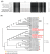
Phylogenetic tree constructed based on the polyprotein amino acid sequences of AmSBV-TW and AcSBV-TW and 54 other SBV strains from the NCBI database. The phylogenetic tree was constructed using the neighbor-joining (NJ) method and 1000 bootstrap replications. The pairwise alignment indicated the deletion patterns in the VP1 region of SBV strains. The round shape symbols indicated AmSBVs with AC genotype (deletion in VP1 region), and the red triangle shape symbols indicated AcSBVs with AM genotype (non-deletion in the VP1 region). red font: The Taiwan strains from this study. *: A note for every 10 bases.
Figure 3
Figure 3
Comparison and phylogenetic analysis of partial VP1 region of AcSBV and AmSBV from Taiwan. (A) The pairwise alignment indicated the 17-anmion deletion presented not in AcSBV but also AmSBV vice versa. (B) Phylogenetic tree construct based on the partial VP-1 amino acid sequences of Taiwan AmSBV and AcSBV, and other 54 SBV strains from NCBI. The phylogenetic tree was constructed using the neighbor-joining (NJ) method and 1000 bootstrap replications. red font: The Taiwan sequences from this study. *: A note for every 10 bases.
Figure 4
Figure 4
Locations of sample collection and the electrophoretic screening of SBV infection-positive samples by RT-PCR with primer set of VP1-F/VP1-R (Supplementary Table S2). The two sample sites for genomic sequencing were located at northern Taiwan; the AcSBV and AmSBV samples were collected in Taipei City and Yilan City, respectively. The black triangle represents the sampling site for detection of partial VP1 region (335 bp) variations in AcSBV and AmSBV in Taiwan. bp = base pair; NC = negative control. The black arrow indicated the signals of SBV positive.

References

    1. Chen Y.P., Pettis J.S., Collins A., Feldlaufer M.F. Prevalence and transmission of honeybee viruses. Appl. Environ. Microbiol. 2006;72:606–611. doi: 10.1128/AEM.72.1.606-611.2006. - DOI - PMC - PubMed
    1. Bailey L., Gibbs A.J., Woods R.D. Sacbrood Virus of the Larval Honey Bee (Apis mellifera Linnaeus) Virology. 1964;23:425–429. doi: 10.1016/0042-6822(64)90266-1. - DOI - PubMed
    1. Baker C.A., Schroeder D.C. The use of RNA-dependent RNA polymerase for the taxonomic assignment of Picorna-like viruses (order Picornavirales) infecting Apis mellifera L. populations. Virol. J. 2008;5:10. doi: 10.1186/1743-422X-5-10. - DOI - PMC - PubMed
    1. Brcak J., Kralik O. On the Structure of the Virus Causing Sacbrood of the Honey Bee. J. Invertebr. Pathol. 1965;20:110–111. doi: 10.1016/0022-2011(65)90166-7. - DOI - PubMed
    1. Zhang J., Feng J., Liang Y., Chen D., Zhou Z.H., Zhang Q., Lu X. Three-dimensional structure of the Chinese Sacbrood bee virus. Sci. China C Life Sci. 2001;44:443–448. doi: 10.1007/BF02879612. - DOI - PubMed