. 2020 Dec 28;9(1):13.
doi: 10.3390/vaccines9010013.

Comparison of Four SARS-CoV-2 Neutralization Assays

Affiliations

Comparison of Four SARS-CoV-2 Neutralization Assays

Lydia Riepler et al. Vaccines (Basel). .

Abstract

Neutralizing antibodies are a major correlate of protection for many viruses including the novel coronavirus SARS-CoV-2. Thus, vaccine candidates should potently induce neutralizing antibodies to render effective protection from infection. A variety of in vitro assays for the detection of SARS-CoV-2 neutralizing antibodies has been described. However, validation of the different assays against each other is important to allow comparison of different studies. Here, we compared four different SARS-CoV-2 neutralization assays using the same set of patient samples. Two assays used replication competent SARS-CoV-2, a focus forming assay and a TCID50-based assay, while the other two assays used replication defective lentiviral or vesicular stomatitis virus (VSV)-based particles pseudotyped with SARS-CoV-2 spike. All assays were robust and produced highly reproducible neutralization titers. Titers of neutralizing antibodies correlated well between the different assays and with the titers of SARS-CoV-2 S-protein binding antibodies detected in an ELISA. Our study showed that commonly used SARS-CoV-2 neutralization assays are robust and that results obtained with different assays are comparable.

Keywords: SARS-CoV-2; neutralization assay; neutralizing antibodies; pseudotype virus.


Conflict of interest statement

The authors declare no conflict of interest.

Figures

Figure 1
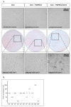
Figure 1
Focus forming neutralization assay. (a) Vero-TMPRSS2 cells were infected with replication competent SARS-CoV-2 virus, fixed after indicated incubation times and stained with the serum of a SARS-CoV-2 convalescent patient. Representative microscopic pictures are shown. (b) Neutralization titers for a set of 11 samples were determined using replication competent SARS-CoV-2 in a focus forming assay. Cells were fixed and stained after 10 h incubation and neutralization titers were determined using ImmunoSpot S6 Ultra-V reader and CTL analyzer BioSpot® 5.0 software (CTL Europe GmbH, Bonn, Germany). The data presented are the result of three independent experiments. Each experiment is represented by a different symbol. Dotted line indicates detection limit for the assay; titers ≤ 1:4 were regarded as negative. (c) Low interassay variability and high reproducibility of the assay was demonstrated by analyzing neutralization titers of a positive control (P3, 1:64 dilution) in 18 independent experiment. Percent neutralization was calculated relative to virus only wells. Shown are individual replicates and mean.
Figure 2
Figure 2
TCID50 neutralization assay. (a) Parental Vero cells, Vero cells expressing TMPRSS2, or Vero cells expressing TMPRSS2 and ACE2 were infected with serial dilutions of SARS-CoV-2. A total of 48 h after infection CPE was analyzed under the microscope and representative microscopic pictures are shown. Additionally, cells were fixed and stained with the serum of a SARS-CoV-2 convalescent patient. Upper panel: noninfected cells; middle panel: immunostaining (10−1 dilution for Vero and Vero-TMPRSS2 cells, 10−5 dilution for Vero-TMPRSS2/ACE2 cells); lower panel: higher magnification of middle panel to evaluate CPE. (b) Neutralization titers for a set of 11 samples were determined using replication competent SARS-CoV-2 in a TCID50 assay. Neutralization titers of three independent experiments are displayed with different symbols. Dotted line indicates detection limit for the assay; titers ≤ 1:4 were regarded as negative.
Figure 3
Figure 3
Vesicular stomatitis virus (VSV)-based neutralization assay. (a) VSVΔG-GFP-S particles were titrated on parental 293T, BHK21, and Vero cells or on cell lines with stable ACE2 overexpression. For 293T and BHK-21 cells two different lentiviral vectors were used for ACE2 overexpression, either with a blasticinin resistance (B) or a hygromycin resistance cassette (H). A total of 16 h after infection, the number of GFP positive cells was determined using an immunospot reader. Shown are mean ± SEM of duplicate samples from one representative of two independent experiments. (b) Neutralization titers for a set of 11 samples were determined using a GFP encoding VSVΔG-S variant. Each sample was analyzed in four independent experiments. Each experiment is represented by a different symbol. Neutralization titers ≤ 1:4 are regarded negative. (c) A positive control (P3 in a 1:64 dilution) was analyzed using the VSVΔG-GFP-S virus on 66 independent plates. Percent neutralization was calculated relative to virus only wells. Shown are individual replicates and mean. (d) Neutralization titers for a set of 11 samples were determined using a luciferase encoding VSVΔG-S variant. Each sample was analyzed in three independent experiments. Each experiment is represented by a different symbol. Dotted line in b + d indicate threshold for neutralizing antibody titers, i.e., titers ≤ 1:4 are regarded as negative. Dotted line in c indicates 50% neutralization.
Figure 4
Figure 4
Lentiviral particle-based neutralization assay. Lentiviral particles encoding GFP were pseudotyped with SARS-CoV-2 S. Neutralization titers for a set of 11 samples were determined. Each sample was analyzed in three independent experiments. Each experiment is represented by a different symbol. Dotted line indicates detection limit for the assay; titers ≤ 1:4 were regarded as negative.
Figure 5
Figure 5
Neutralization titers for the different assays correlate well with each other and with binding antibody titers. For neutralization assays mean neutralizing titers of three (focus forming, TCID50, and lentiviral particle assay) or four independent experiments (VSV assay) were calculated. (a) Correlation of neutralizing antibody titers and binding antibodies (anti-S IgA, anti-S IgG, and anti-N IgG) were determined. (b) Neutralizing antibody titers determined in each assay were correlated to titers in all three other neutralizing antibody assays. Dotted lines indicate detection limit for the different assays; neutralization assay ≤ 1:4 negative, anti-S IgA and anti-S IgG OD < 0.8 negative, OD 0.8–1.1 borderline, OD > 1.1 positive, anti-N IgG RLU > 1.4 positive. Some of the dots in the blots represent multiple samples.

References

    1. World Health Organization WHO Coronavirus Disease (COVID-19) Dashboard. [(accessed on 22 December 2020)]; Available online: https://covid19.who.int.
    1. FIND SARS-CoV-2 Diagnostic Pipeline. [(accessed on 22 December 2020)]; Available online: https://www.finddx.org/covid-19/pipeline/
    1. Meyer B., Torriani G., Yerly S., Mazza L., Calame A., Arm-Vernez I., Zimmer G., Agoritsas T., Stirnemann J., Spechbach H., et al. Validation of a commercially available SARS-CoV-2 serological immunoassay. Clin. Microbiol. Infect. 2020;26:1386–1394. doi: 10.1016/j.cmi.2020.06.024. - DOI - PMC - PubMed
    1. Grzelak L., Temmam S., Planchais C., Demeret C., Tondeur L., Huon C., Guivel-Benhassine F., Staropoli I., Chazal M., Dufloo J., et al. A comparison of four serological assays for detecting anti-SARS-CoV-2 antibodies in human serum samples from different populations. Sci. Transl. Med. 2020;12:eabc3103. doi: 10.1126/scitranslmed.abc3103. - DOI - PMC - PubMed
    1. Zhao J., Yuan Q., Wang H., Liu W., Liao X., Su Y., Wang X., Yuan J., Li T., Li J., et al. Antibody responses to SARS-CoV-2 in patients of novel coronavirus disease 2019. Clin. Infect. Dis. 2020;71:2027–2034. doi: 10.1093/cid/ciaa344. - DOI - PMC - PubMed