. 2020 Dec 28;10(1):32.
doi: 10.3390/cells10010032.

Cyclic Stretch of Either PNS or CNS Located Nerves Can Stimulate Neurite Outgrowth

Affiliations

Cyclic Stretch of Either PNS or CNS Located Nerves Can Stimulate Neurite Outgrowth

Vasileios Kampanis et al. Cells. .

Abstract

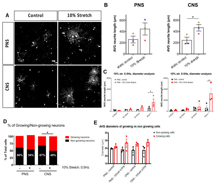
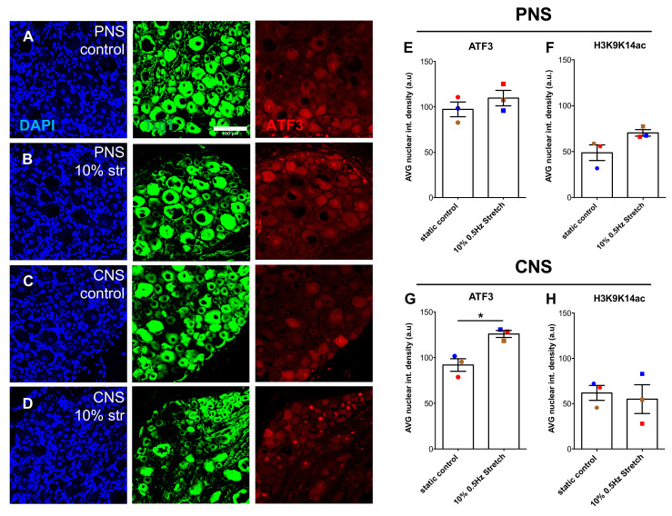
The central nervous system (CNS) does not recover from traumatic axonal injury, but the peripheral nervous system (PNS) does. We hypothesize that this fundamental difference in regenerative capacity may be based upon the absence of stimulatory mechanical forces in the CNS due to the protective rigidity of the vertebral column and skull. We developed a bioreactor to apply low-strain cyclic axonal stretch to adult rat dorsal root ganglia (DRG) connected to either the peripheral or central nerves in an explant model for inducing axonal growth. In response, larger diameter DRG neurons, mechanoreceptors and proprioceptors showed enhanced neurite outgrowth as well as increased Activating Transcription Factor 3 (ATF3).

Keywords: CNS; DRG; PNS; cyclic stretch; mechanical loading; neurite outgrowth.


Conflict of interest statement

The authors declare no conflict of interest. The funders had no role in the design of the study; in the collection, analyses, or interpretation of data; in the writing of the manuscript, or in the decision to publish the results.

Figures

Figure 1
Figure 1
In-house built setup for the mechanical stretch of nerve explants. (A) The controller is connected to GalilTools software and the oscillator (D,E). (B) The incubator chamber keeping constant culture conditions. (C) The gas mixer analyzing the air constitution of the incubator chamber, 5% CO2. (D) The bioreactor (F) is mounted on the oscillator (E). (E) The oscillator is connected to the controller (A) and the bioreactor (F) inside the incubator. (F) Bioreactor. Two metal plates (a and b) are built on a metal rail, and poly-ether-ether-ketone (PEEK) bars are fixed on them by screws and kept in a metal boat filled with culture medium. Metal plate b is connected to the oscillator, moving back and forth, while the other plate is fixed to the stage and the metal rail.
Figure 2
Figure 2
Peripheral nervous system (PNS) and central nervous system (CNS) nerve-dorsal root ganglia (DRG) explants. (A) The L4-L5 DRG are either connected to the sciatic nerve or continue independently along their dorsal roots into the spinal column. Two groups were prepared with either the central nerves connected to their respective L4/L5 DRG or the sciatic nerve connected to the L4/L5 DRG. (B) Explant nerves were glued on two PEEK bars facing towards each other, with the DRG sitting on one bar and the end of the nerve(s) seated on the other bar with a 1 cm distance between them. With the 10% cyclic movement of plate B (lower metal plate), 1 mm dislocation was achieved.
Figure 3
Figure 3
Ex vivo mechanical stretch enhances neurite outgrowth. Three hours following a mechanical stretch of either the sciatic nerve connected to L4-L5 DRG or connected to their respective central nerves, DRG were dissected and cultured for 18 h on laminin substrates. (A) Immunohistochemical staining of βIII-tubulin (Tuj1) was used to trace all neurites of the DRG. Scale bar, 100 μm, (B) 10% mechanical stretch with a frequency of 0.5 Hz enhanced neurite outgrowth of DRG derived from a CNS explant but not from a PNS explant. Paired two-tailed Student t-test, mean ± SEM of paired independent experiments indicated by the colors, * p < 0.05, (C) Diameter analysis of DRG outgrowth comparing the static controls to the stretched explants shows the largest neurons responding to stretch. Two-way ANOVA with post-hoc Sidak’s multiple comparison test, mean ± SEM, * p < 0.05, ** p < 0.01. (D) Quantification of growing vs. non-growing neurons in each group showed that non-growing neurons decreased in the CNS group upon the stretch. Two-way ANOVA with post-hoc Sidak’s multiple comparison test, * p < 0.05. (E) Examination of the average size of growing versus non-growing neurons shows non-growing neurons to be consistently smaller diameters than growing neurons.
Figure 4
Figure 4
20% 0.5 Hz stretch has a negative impact on neurite outgrowth of DRG in vitro. (A) Representative photomicrographs (10×) of cultured rat DRG following 20% 0.5 Hz stretch or no stretch and stained for βΙΙΙ-tubulin (Tuj1). Scale bar, 200 μm. (B) Neurite length analysis of DRG cultured after 20% 0.5 Hz stretch of PNS-nerve explant compared to static control. Stretch appeared to have a negative impact on the neurite outgrowth, although not statistically significant. Paired two-tailed Student’s t-test, mean ± SEM of paired independent experiments indicated by the colors. (C) Diameter analysis showing a statistically significant reduction in the outgrowth of large-diameter neurons after a 20% stretch of the PNS-nerve explant. Two-way ANOVA with post-hoc Sidak’s multiple comparison test, mean ± SEM, ** p < 0.01, (D) Neurite length analysis of DRG cultured after 20% 0.5 Hz stretch of CNS-nerve explant compared to static control. Stretch appeared to have a negative impact on the neurite outgrowth of these cells as well, although not statistically significant due to high variability. Paired two-tailed Student’s t-test, mean ± SEM of paired independent experiments indicated by the colors. (E) Diameter analysis showing a reduction trend in the outgrowth of all DRG populations after 20% stretch of the CNS-nerve explant. Two-way ANOVA with post-hoc Sidak’s multiple comparison test, mean ± SEM, ** p < 0.01.
Figure 5
Figure 5
3 h of 10% stretch does not significantly alter the microarchitecture of the nerve, although 20% stretch does. (AF) Photomicrographs (10×) of H&E staining of sciatic nerves (AC) and central nerves (DF). Nerves were fixed and dissected longitudinally after either no stretch (A,D) or mechanical loading of 10% (B,E) or 20% (C,F). Arrows in (A) indicate the epineurium (Ep), perineurium (P), and endoneurium (En). The epineurium, specific to the PNS, is densely formed in the static controls but begins to unravel upon 20% stretch. The perineurium, a protective sheath covering nerve fascicles, was found to slowly thin with increasing amounts of stretch, with some tears in the 20% samples. Notably, the naturally occurring nerve undulation observed in the static control (Bands of Fontana) is progressively lost upon the mechanical stretch. Scale bar, 500 μm.
Figure 6
Figure 6
NF200+ DRG neurons respond to mechanical stretch. (A) Immunocytochemical analysis of DRG stained for NF200 after either no stretch or mechanical stretch (10% 0.5 Hz). Scale bar, 200 μm (B,D) Neurite length analysis of NF200+ PNS (B) and CNS (D) DRG upon 10% 0.5 Hz mechanical stretch. Paired two-tail Student t-test, mean ± SEM of paired independent experiments indicated by the colors. (C,E) Diameter analysis of PNS (C) and CNS (E) DRG outgrowth comparing the static controls to the stretched explants shows that after peripheral stretch 30–40 µm-diameter neurons increase their neurite outgrowth. Two-way ANOVA with post-hoc Sidak’s multiple comparison test, mean ± SEM, ** p < 0.01.
Figure 7
Figure 7
TrkC+ but not CGRP+ DRG neurons respond after mechanical stretch. (A) Immunocytochemical analysis of DRG stained either for TrkC (green) or CGRP (red) after either no stretch or mechanical stretch (10% 0.5 Hz). Scale bar, 200 μm (B) Neurite length analysis of TrkC+ DRG upon mechanical stretch with an amplitude of either 10% or 20% and a frequency of 0.25 Hz or 0.5 Hz. (C) Neurite length analysis of CGRP+ DRG upon mechanical stretch with an amplitude of either 10% or 20% and a frequency of 0.25 Hz or 0.5 Hz. Two-way ANOVA, with post-hoc Sidak’s multiple comparison test, mean ± SEM, * p < 0.05.
Figure 8
Figure 8
Within 3 h of 10% mechanical stretch, ATF3 nuclear DRG levels increased in the CNS DRG-nerve explant. (AD) Representative immunofluorescence photomicrographs (20×) of DRG sectioned either 3 h post- no stimulation (A) or 10% mechanical stretch (B) of PNS DRG-nerve explants and no stimulation (C) or 10% mechanical stretch (D) of CNS DRG-nerve explants. DRG were stained for DAPI (blue), βIII-tubulin (Tuj1) (green), and ATF3 (red). Scale bar, 100 μm. (E,G) Nuclear intensity density analysis of ATF3 in DRG derived from PNS DRG-nerve explants (E) and CNS DRG-nerve explants (G). Paired two-tailed Student t-test, mean ± SEM of paired independent experiments indicated by the colors, * p < 0.05. (F,H) Nuclear intensity density analysis of H3K9K14ac in DRG derived from PNS DRG-nerve explants (F) and CNS DRG-nerve explants (H). Paired two-tailed Student t-test, mean ± SEM of paired independent experiments indicated by the colors.

Similar articles

Cited by

References

    1. Mammoto T., Ingber D.E. Mechanical control of tissue and organ development. Development. 2010;137:1407–1420. doi: 10.1242/dev.024166. - DOI - PMC - PubMed
    1. Franze K. The mechanical control of nervous system development. Development. 2013;140:3069–3077. doi: 10.1242/dev.079145. - DOI - PubMed
    1. LeGoff L., Lecuit T. Mechanical forces and growth in animal tissues. Cold Spring Harb. Perspect. Biol. 2016;8:1–17. doi: 10.1101/cshperspect.a019232. - DOI - PMC - PubMed
    1. Geffeney S.L., Goodman M.B. How We Feel: Ion Channel Partnerships that Detect Mechanical Inputs and Give Rise to Touch and Pain Perception. Neuron. 2012;74:609–619. doi: 10.1016/j.neuron.2012.04.023. - DOI - PMC - PubMed
    1. Proske U., Gandevia S.C. The proprioceptive senses: Their roles in signaling body shape, body position and movement, and muscle force. Physiol. Rev. 2012;92:1651–1697. doi: 10.1152/physrev.00048.2011. - DOI - PubMed

Publication types

Substances

LinkOut - more resources