. 2020 Dec 28;22(1):217.
doi: 10.3390/ijms22010217.

Kaempferol Inhibits Zearalenone-Induced Oxidative Stress and Apoptosis via the PI3K/Akt-Mediated Nrf2 Signaling Pathway: In Vitro and In Vivo Studies

Affiliations

Kaempferol Inhibits Zearalenone-Induced Oxidative Stress and Apoptosis via the PI3K/Akt-Mediated Nrf2 Signaling Pathway: In Vitro and In Vivo Studies

Peramaiyan Rajendran et al. Int J Mol Sci. .

Abstract

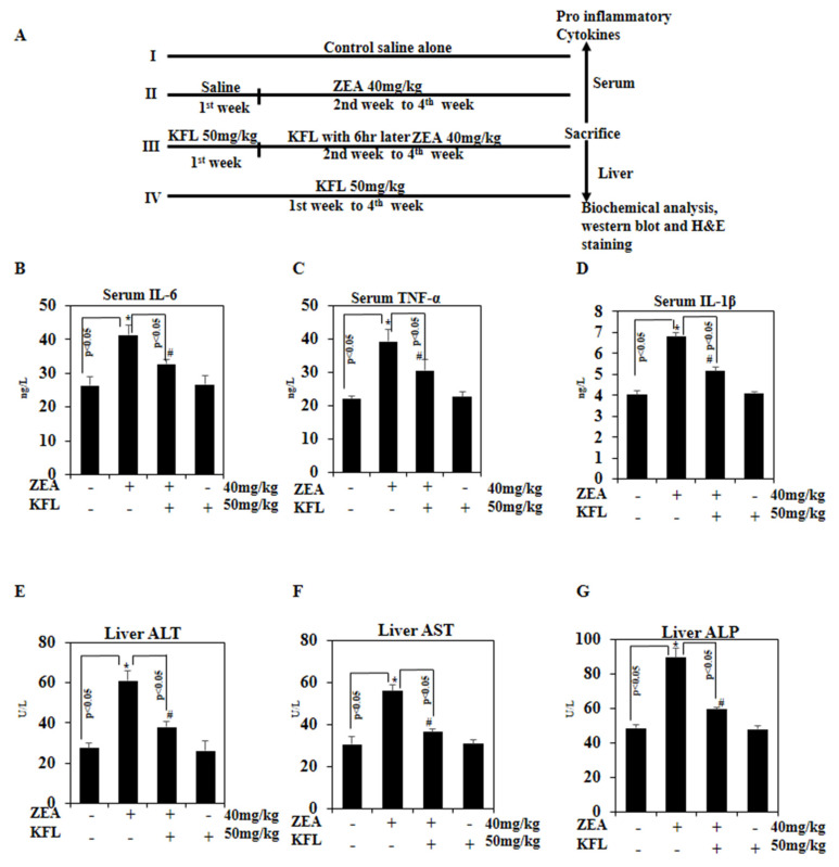
In this study, kaempferol (KFL) shows hepatoprotective activity against zearalenone (ZEA)-induced oxidative stress and its underlying mechanisms in in vitro and in vivo models were investigated. Oxidative stress plays a critical role in the pathophysiology of various hepatic ailments and is normally regulated by reactive oxygen species (ROS). ZEA is a mycotoxin known to exert toxicity via inflammation and ROS accumulation. This study aims to explore the protective role of KFL against ZEA-triggered hepatic injury via the PI3K/Akt-regulated Nrf2 pathway. KFL augmented the phosphorylation of PI3K and Akt, which may stimulate antioxidative and antiapoptotic signaling in hepatic cells. KFL upregulated Nrf2 phosphorylation and the expression of antioxidant genes HO-1 and NQO-1 in a dose-dependent manner under ZEA-induced oxidative stress. Nrf2 knockdown via small-interfering RNA (siRNA) inhibited the KFL-mediated defence against ZEA-induced hepatotoxicity. In vivo studies showed that KFL decreased inflammation and lipid peroxidation and increased H2O2 scavenging and biochemical marker enzyme expression. KFL was able to normalize the expression of liver antioxidant enzymes SOD, CAT and GSH and showed a protective effect against ZEA-induced pathophysiology in the livers of mice. These outcomes demonstrate that KFL possesses notable hepatoprotective roles against ZEA-induced damage in vivo and in vitro. These protective properties of KFL may occur through the stimulation of Nrf2/HO-1 cascades and PI3K/Akt signaling.

Keywords: Nrf2; PI3K/Akt; apoptosis; hepatotoxicity; kaempferol; zearalenone.


Conflict of interest statement

The authors declare no conflict of interest.

Figures

Figure 1
Figure 1
Impact of zearalenone (ZEA) and kaempferol (KFL) on viability. (A) Chemical structure of kaempferol. (B) HepG2 cells were added with the indicated dosages of ZEA for 24 h and 48 h, and then viability was determined through MTT assay. (C) Cytotoxic effect of KFL on HepG2 cells. Cells were exposed to different concentrations of KFL for 24 h. (D) KFL protects the cytotoxic effect of ZEA (24 h) as analyzed by MTT assay. Data are represented as the mean ± SD of triplicate values (n = 3), and * p < 0.05 represents significant difference in comparison to the control group. # p < 0.05 represents significant variations compared the ZEA alone and KFL with ZEA treatment groups.
Figure 2
Figure 2
KFL induces nuclear factor-erythroid 2-related factor 2-dependent haem oxygenase-1 (HO-1) expression in ZEA-treated HepG2 cells. (A) Cells were incubated with various concentrations of ZEA for 24 h, and equal amounts of whole-cell lysate were exposed to SDS-PAGE. Membranes probed with anti-NQO1, anti-HO-1 and anti-Nrf2 antibodies. (B) Cells were treated with KFL (50 μM) and/or ZEA (40 μM) for 24 h, and the Western blotting results show the effects of KFL on the protein levels of HO-1, NQO-1, and Nrf2 in the cytosolic and nuclear fractions. The relative ratios of expression in the Western blotting results are demonstrated below each of the results as values relative to actin expression. Data are represented as the mean ± SD of triplicate values (n = 3), and * p < 0.05 represents significant variations compared with the control. # p < 0.05 represents remarkable variations compared the ZEA alone and KFL with ZEA treatment groups.
Figure 3
Figure 3
KFL up-regulates the antioxidant markers Nrf2, NQO-1 and HO-1 in HepG2 cells. (A) Nrf2, NQO-1 and HO-1 expression analyzed by RT-PCR. (B) The effects of inhibiting Nrf2 by siRNA transfection were studied by protein analysis. Cells were transiently transfected with Nrf2 siRNA at 100 pM per well in 6-well plates for 24 h, followed by treatment with ZEA and/or KFL for 24 h. SDS-PAGE was used to detect the expression of Nrf2 and HO-1 proteins. Data are represented as the mean ± SD of triplicate values (n = 3), and * p < 0.05 denotes remarkable variations as compared to the control. # p < 0.05 denotes remarkable variations compared the ZEA alone and KFL with ZEA treatment groups.
Figure 4
Figure 4
KFL triggers the PI3K/Akt pathway in ZEA-induced HepG2 cells. (A) Cells were treated with KFL (10, 25 and 50 µM) followed by ZEA (40 µM) for 24 h. After the treatment, whole-cell lysates were exposed to Western blotting with anti-pPI3K and anti-pAkt antibodies. Total PI3K and Akt levels were measured as loading controls. (B) Cells were pre-treated with a PI3K/Akt inhibitor (LY294002, 30 μM) for 2 h, followed by KFL (50 μM) and/or ZEA (40 μM) for 24 h. Western blot was performed to detect the pAkt, HO-1 and Nrf2 levels by anti-pAkt, anti-HO-1 and anti-Nrf2 abs. Data are represented as the mean ± SD of triplicate values (n = 3), and * p < 0.05 represents noteworthy discrepancies compared with the control. # p < 0.05 represents significant variations compared with the ZEA alone and KFL with ZEA treatment groups.
Figure 5
Figure 5
KFL repressed DNA damage and apoptosis induced by ZEA in HepG2 cells (A) γ-H2AX nuclear protein expression analyzed and γ-H2AX normalized to Lamin B1. (B) DNA fragmentation (C) HepG2 cells were treated with the KFL and/or ZEA for 24 h. Expression of BCL2, cleaved caspase-3 (clvd Caspase3) and cleaved PARP (clvd PARP) protein was assessed by Western blotting. (D) KFL protected against ZEA-induced apoptosis (24 h), as indicated by TUNEL assay. Data are represented as the mean ± SD of triplicate values (n = 3), and * p < 0.05 represents significant variations compared with the control. # p < 0.05 represents significant variations as compared to ZEA alone and KFL with ZEA treatment groups.
Figure 6
Figure 6
Effect of KFL on pro-inflammatory cytokines in ZEA-treated mice. (A) Schematic of the animal experimental method. (BD) ELISA was used to quantity the levels of the pro-inflammatory cytokines (B) IL-6, (C) TNF-α and (D) IL-1β in mouse serum. (EG) The level of liver markers was analyzed by ELISA: (E) ALT, (F) AST and (G) ALP. Data are expressed as the mean values ± SD of independent experiments (n = 6); * p < 0.05 represents significant variations in the ZEA alone group compared with the control group. # p < 0.05 represents significant variations compared the ZEA alone and KFL with ZEA treatment groups.
Figure 7
Figure 7
Effect of KFL on ZEA-induced ROS accumulation and antioxidant systems in mouse livers. (A) H2O2, (B) LPO, (C) SOD, (D) CAT and (E) GSH. ELISA kits were used based on the manufacturer’s directions (units: H2O2, µm/g protein; LPO, nM of MDA released/mg protein; SOD, CAT and GSH, U/mg protein). Data are expressed as the mean values ± SD of independent experiments (n = 6). * Significant in the ZEA alone group compared with the control group (p < 0.05). # p < 0.05 represents significant variations compared the ZEA alone and KFL with ZEA treatment groups.
Figure 8
Figure 8
Effect of KFL on histopathological changes in control and experimental animals. (A) Microscopic pictures of mouse liver tissue (H&E, 400×). Representative images of H&E staining of liver tissue from mice after treatment with ZEA alone or with both ZEA and KFL: panel (a)—control, hepatocyte cords (Hc), clearly visible nuclei of hepatocytes, central vein (C) and normal hepatocytes visible panel (b,c)—ZEA alone, (B)—Circle signifies infiltrated inflammatory cells, panel (c)—KFL with ZEA, lesser sinusoidal congestion and some infiltrated inflammatory cells and panel (d)—KFL alone, marked arrow showed the normal hepatocytes and decrease of sinusoidal congestion, reduction in hepatocytes swelling exhibit recovery and retention of tissue integrity. (B) The histopathological grade values of the hepatocyte necrosis and inflammatory cell infiltration into the liver at the end of the experiment were measured (n = 6). (C) Inflections in pPI3K, pAkt, Nrf2 and Bcl2 proteins in albino mice. β-Actin was used as the internal control. Data are articulated as the mean values ± SD of independent experiments (n = 6). * Significant in the ZEA alone group compared with the control group (p < 0.05). # p < 0.05 represents significant variations compared to the ZEA alone and KFL with ZEA treatment groups.

References

    1. Rajput S.A., Sun L., Zhang N.-Y., Khalil M.M., Ling Z., Chong L., Wang S., Rajput I.R., Bloch D.M., Khan F.A. Grape seed proanthocyanidin extract alleviates aflatoxinB1-induced immunotoxicity and oxidative stress via modulation of NF-κB and Nrf2 signaling pathways in broilers. Toxins. 2019;11:23. doi: 10.3390/toxins11010023. - DOI - PMC - PubMed
    1. Fernández-Cruz M.L., Mansilla M.L., Tadeo J.L. Mycotoxins in fruits and their processed products: Analysis, occurrence and health implications. J. Adv. Res. 2010;1:113–122. doi: 10.1016/j.jare.2010.03.002. - DOI
    1. Lee H.-J., Park J.-H., Oh S.-Y., Cho D.-H., Kim S., Jo I. Zearalenone-induced interaction between PXR and Sp1 increases binding of Sp1 to a promoter site of the eNOS, decreasing its transcription and NO production in BAECs. Toxins. 2020;12:421. doi: 10.3390/toxins12060421. - DOI - PMC - PubMed
    1. Yoon J.E., Lee K.Y., Seok J.S., Cheng W.N., Kwon H.C., Jeong C.H., Han S.G. Zearalenone induces endoplasmic reticulum stress and modulates the expression of phase I/II enzymes in human liver cells. Toxins. 2020;12:2. doi: 10.3390/toxins12010002. - DOI - PMC - PubMed
    1. Skiepko N., Przybylska-Gornowicz B., Gajęcka M., Gajęcki M., Lewczuk B. Effects of deoxynivalenol and zearalenone on the histology and ultrastructure of pig liver. Toxins. 2020;12:463. doi: 10.3390/toxins12070463. - DOI - PMC - PubMed

MeSH terms

LinkOut - more resources