Review
. 2020 Dec 28;22(1):238.
doi: 10.3390/ijms22010238.

Hold on or Cut? Integrin- and MMP-Mediated Cell-Matrix Interactions in the Tumor Microenvironment

Affiliations
Review

Hold on or Cut? Integrin- and MMP-Mediated Cell-Matrix Interactions in the Tumor Microenvironment

Stephan Niland et al. Int J Mol Sci. .

Abstract

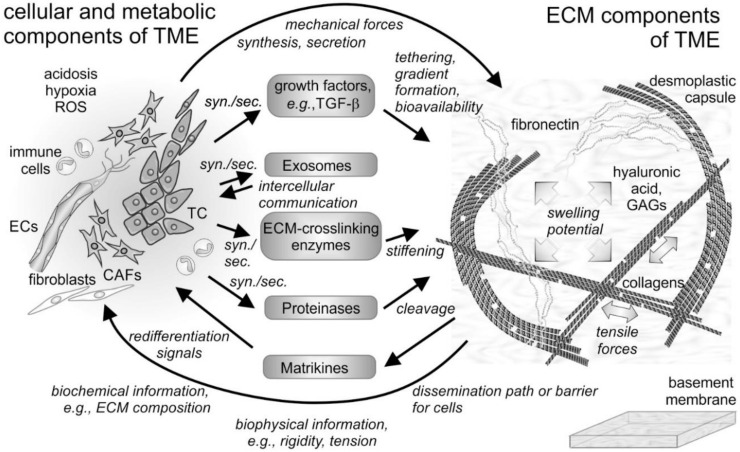
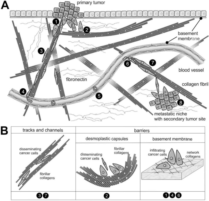
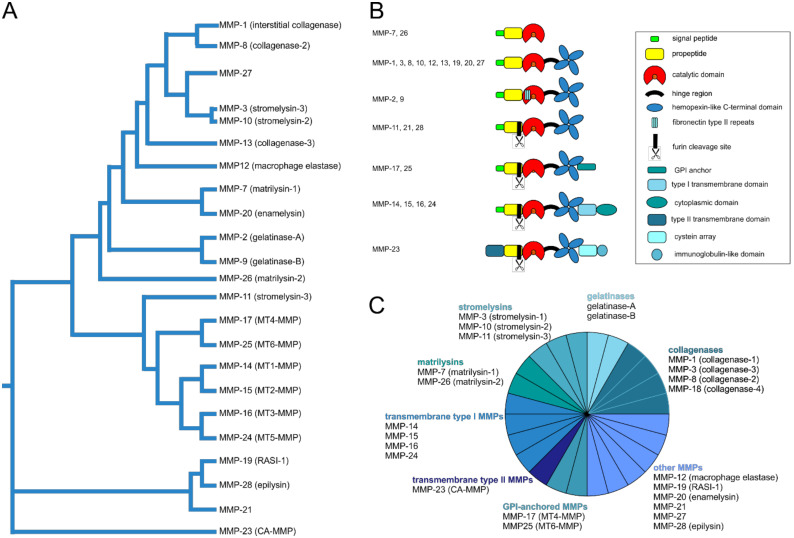
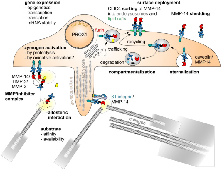
The tumor microenvironment (TME) has become the focus of interest in cancer research and treatment. It includes the extracellular matrix (ECM) and ECM-modifying enzymes that are secreted by cancer and neighboring cells. The ECM serves both to anchor the tumor cells embedded in it and as a means of communication between the various cellular and non-cellular components of the TME. The cells of the TME modify their surrounding cancer-characteristic ECM. This in turn provides feedback to them via cellular receptors, thereby regulating, together with cytokines and exosomes, differentiation processes as well as tumor progression and spread. Matrix remodeling is accomplished by altering the repertoire of ECM components and by biophysical changes in stiffness and tension caused by ECM-crosslinking and ECM-degrading enzymes, in particular matrix metalloproteinases (MMPs). These can degrade ECM barriers or, by partial proteolysis, release soluble ECM fragments called matrikines, which influence cells inside and outside the TME. This review examines the changes in the ECM of the TME and the interaction between cells and the ECM, with a particular focus on MMPs.

Keywords: extracellular matrix; integrins; matrikines; matrix metalloproteinases; tumor microenvironment.


Conflict of interest statement

The authors declare that they have no conflict of interest.

Figures

Figure 1
Figure 1
Cellular and non-cellular components of the tumor microenvironment (TME) and their interplay. In addition to the tumor cells (TCs), fibroblasts and their derivatives, the cancer-associated fibroblasts (CAFs), as well as ingrowing endothelial cells (ECs) and infiltrating immune cells are the cellular components of the tumor microenvironment (TME). The cells synthesize and secrete (syn./sec.) not only the extracellular matrix (ECM) components, but also growth factors, exosomes, and ECM-modifying enzymes, such as proteinases. Both cells and their secretion products interact with the fibrillar and non-fibrillar components of the ECM. Hyaluronic acid and glycosaminoglycan (GAG)-chains of proteoglycans increase the swelling potential of the interstitial space, which is counterbalanced by the tensile force-bearing fibrils of collagens, elastin and fibronectin. Modified by tethered growth factors, by crosslinking and cleaving enzymes, and by contractile forces, the ECM and its fragments with cytokine-like functions (matrikines) influence the cells within the TME in various ways. ROS, reactive oxygen species.
Figure 2
Figure 2
The supramolecular structure of collagen as a substrate or impediment of cancer cell dissemination: (A) The metastatic cascade of malignant carcinoma cells includes the penetration of the epithelial basement membrane (❶); Solid tumors within the stroma tissue are often surrounded by a desmoplastic collagen capsule, which impedes cancer cell migration (❷); Through the interstitial stroma, cancer cells utilize collagen-rich fibrils to quickly reach a nearby blood vessel (❸); There, they intravasate through the endothelial basement membrane (❹); When blood-borne, they float with the blood stream, mostly sheltered by platelets (❺); To extravasate, they attach to the vessel wall of a distant organ and again breach the endothelial basement membrane (❻); Moving quickly along collagen-rich fibrils (❼), they reach the metastatic niche (❽), where they grow into a metastasis. (B) The different dissemination-supporting and impeding functions of the collagen suprastructures are highlighted for the different steps of the metastatic cascade. The cancer cells experience, particularly by integrins, the ECM as dissemination-supporting tracks and channels, especially if migration and fibrils point into the same direction and if the fibril network is not too dense. In contrast, the dense array of collagen fibrils in desmoplastic capsules and of network-forming collagens within the basement membrane impedes cancer cell progression and requires the use matrix-metalloproteinases. Moreover, the orientation of collagen fibrils within the desmoplastic capsule is mostly perpendicular to the direction of dissemination [28].
Figure 3
Figure 3
Phylogenetic and functional relationship of human matrix metalloproteinases (MMPs). In addition to the numbering of the MMPs, which corresponds to the order of their discovery, the MMPs can be ordered according to their sequence similarity [154,155] (A). Regarding their domain organization (B) and substrate specificity they can be assigned to different groups within the MMP family (C): soluble collagenases, gelatinases, stromelysins, matrilysins, membrane-anchored transmembrane type I and type II as well as glycosylphosphatidylinositol (GPI)-anchored MMPs, and other MMPs [152]. All MMPs except MMP-23 have a propeptide with an N-terminal signal sequence. To activate an MMP, this propeptide has to be cleaved off in order to make a zinc ion in the active site of the catalytic domain, which also holds calcium ions, accessible through a cysteine switch. CA-MMP, cysteine array matrix metalloproteinase; RASI-1, rheumatoid arthritis synovium inflamed-1.
Figure 4
Figure 4
MMP-14 is regulated by various mechanisms. The enzymatic activity of MMP-14 is regulated by various mechanisms: at the level of gene expression, there are epigenetic [172], transcriptional, and posttranscriptional regulatory mechanisms, such as the cotranslational cleavage of its signal sequence at the endoplasmatic reticulum, the furin-mediated removal of its self-inhibitory prodomain within the Golgi compartment [174], the O-glycosylation of protease-sensitive linker regions, and the phosphorylation as well as palmitoylation of its cytoplasmic domain [175,176], with the transcription factor PROX1 playing an important role [157]. Zymogen activation [156], compartmentalization, surface deployment and internalization [177,178], sorting into lipid rafts [179], homodimerization and interaction with other proteins, such as TIMPs [180], integrin β1 [181], and substrates, as well as shedding [182,183] are closely interlinked.

References

    1. He Q., Chen J., Yan J., Cai S., Xiong H., Liu Y., Peng D., Mo M., Liu Z. Tumor microenvironment responsive drug delivery systems. Asian J. Pharm. Sci. 2020;15:416–448. doi: 10.1016/j.ajps.2019.08.003. - DOI - PMC - PubMed
    1. Emon B., Bauer J., Jain Y., Jung B., Saif T. Biophysics of Tumor Microenvironment and Cancer Metastasis—A Mini Review. Comput. Struct. Biotechnol. J. 2018;16:279–287. doi: 10.1016/j.csbj.2018.07.003. - DOI - PMC - PubMed
    1. Mohammadi H., Sahai E. Mechanisms and impact of altered tumour mechanics. Nat. Cell Biol. 2018;20:766–774. doi: 10.1038/s41556-018-0131-2. - DOI - PubMed
    1. Iozzo R.V., Schaefer L. Proteoglycan form and function: A comprehensive nomenclature of proteoglycans. Matrix Biol. 2015;42:11–55. doi: 10.1016/j.matbio.2015.02.003. - DOI - PMC - PubMed
    1. Theocharis A.D., Skandalis S.S., Gialeli C., Karamanos N.K. Extracellular matrix structure. Adv. Drug Deliv. Rev. 2016;97:4–27. doi: 10.1016/j.addr.2015.11.001. - DOI - PubMed