

Cognitive impact of COVID-19: looking beyond the short term
- PMID: 33380345
- PMCID: PMC7772800
- DOI: 10.1186/s13195-020-00744-w
Cognitive impact of COVID-19: looking beyond the short term
Abstract
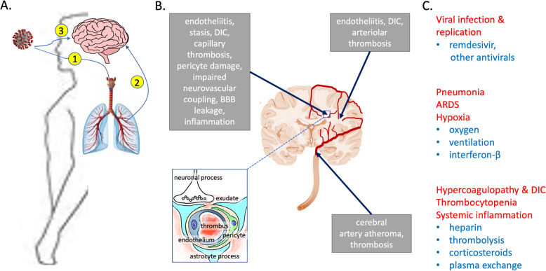
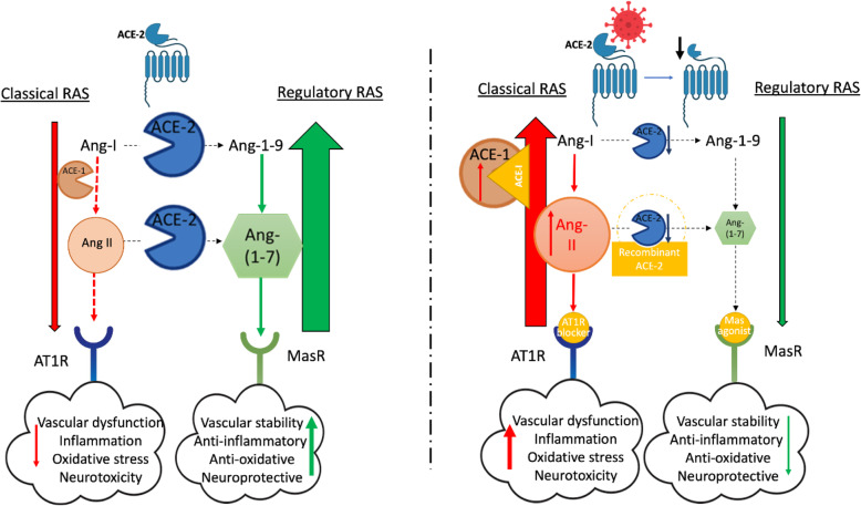
COVID-19 is primarily a respiratory disease but up to two thirds of hospitalised patients show evidence of central nervous system (CNS) damage, predominantly ischaemic, in some cases haemorrhagic and occasionally encephalitic. It is unclear how much of the ischaemic damage is mediated by direct or inflammatory effects of virus on the CNS vasculature and how much is secondary to extracranial cardiorespiratory disease. Limited data suggest that the causative SARS-CoV-2 virus may enter the CNS via the nasal mucosa and olfactory fibres, or by haematogenous spread, and is capable of infecting endothelial cells, pericytes and probably neurons. Extracranially, SARS-CoV-2 targets endothelial cells and pericytes, causing endothelial cell dysfunction, vascular leakage and immune activation, sometimes leading to disseminated intravascular coagulation. It remains to be confirmed whether endothelial cells and pericytes in the cerebral vasculature are similarly targeted. Several aspects of COVID-19 are likely to impact on cognition. Cerebral white matter is particularly vulnerable to ischaemic damage in COVID-19 and is also critically important for cognitive function. There is accumulating evidence that cerebral hypoperfusion accelerates amyloid-β (Aβ) accumulation and is linked to tau and TDP-43 pathology, and by inducing phosphorylation of α-synuclein at serine-129, ischaemia may also increase the risk of development of Lewy body disease. Current therapies for COVID-19 are understandably focused on supporting respiratory function, preventing thrombosis and reducing immune activation. Since angiotensin-converting enzyme (ACE)-2 is a receptor for SARS-CoV-2, and ACE inhibitors and angiotensin receptor blockers are predicted to increase ACE-2 expression, it was initially feared that their use might exacerbate COVID-19. Recent meta-analyses have instead suggested that these medications are protective. This is perhaps because SARS-CoV-2 entry may deplete ACE-2, tipping the balance towards angiotensin II-ACE-1-mediated classical RAS activation: exacerbating hypoperfusion and promoting inflammation. It may be relevant that APOE ε4 individuals, who seem to be at increased risk of COVID-19, also have lowest ACE-2 activity. COVID-19 is likely to leave an unexpected legacy of long-term neurological complications in a significant number of survivors. Cognitive follow-up of COVID-19 patients will be important, especially in patients who develop cerebrovascular and neurological complications during the acute illness.
Keywords: Angiotensin receptor blockers; Angiotensin-converting enzyme inhibitors; Angiotensin-converting enzyme-2; COVID-19; Cognitive impairment; Dementia; SARS-CoV-2; Stroke; White matter ischaemia.
Conflict of interest statement
The authors declare no competing interests.
Figures
References
Publication types
MeSH terms
LinkOut - more resources
Full Text Sources
Medical
Miscellaneous
