. 2021 Feb 15;206(4):797-806.
doi: 10.4049/jimmunol.2000091. Epub 2020 Dec 30.

Inhibition of Efferocytosis by Extracellular CIRP-Induced Neutrophil Extracellular Traps

Affiliations

Inhibition of Efferocytosis by Extracellular CIRP-Induced Neutrophil Extracellular Traps

Kehong Chen et al. J Immunol. .

Abstract

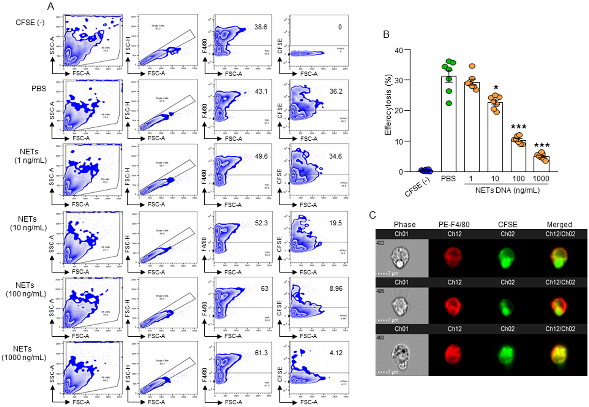
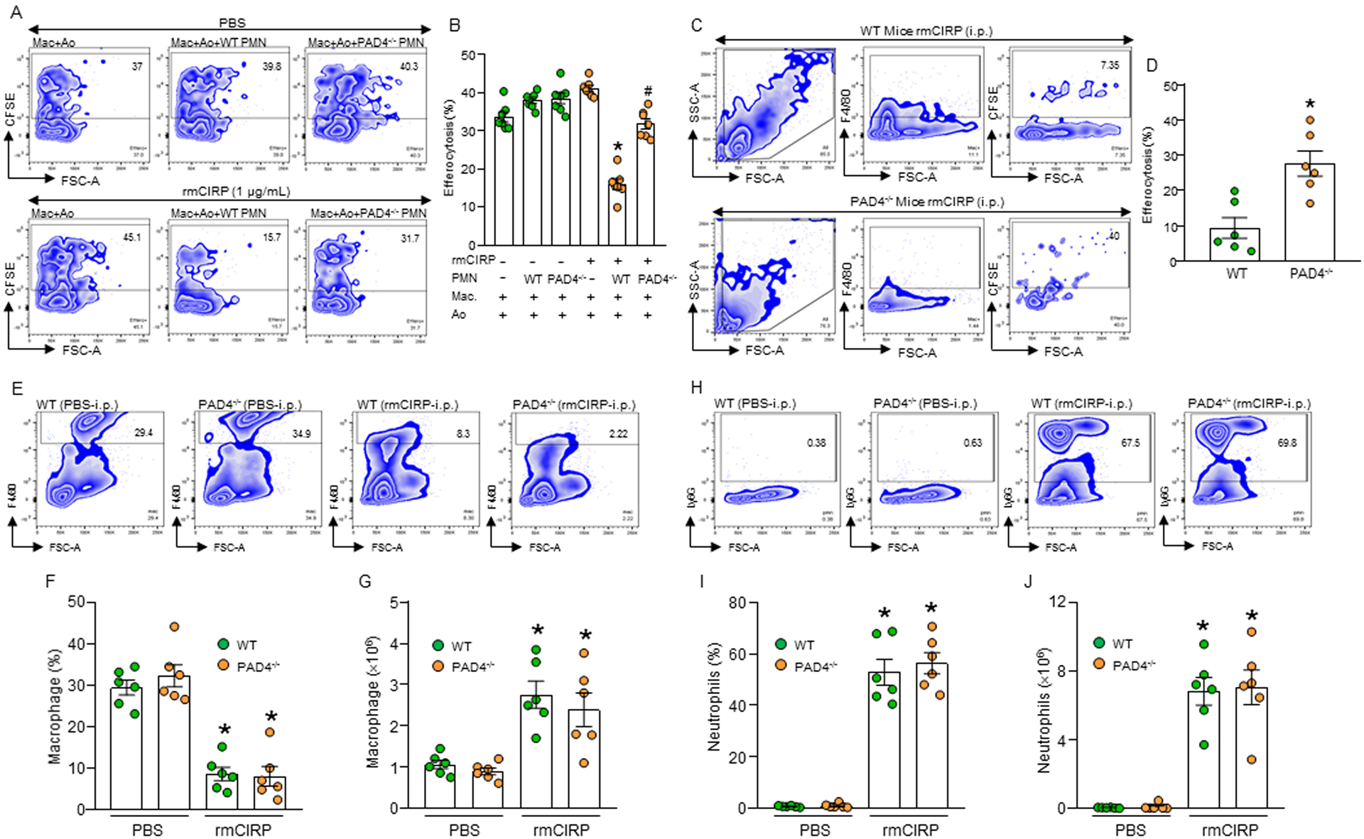
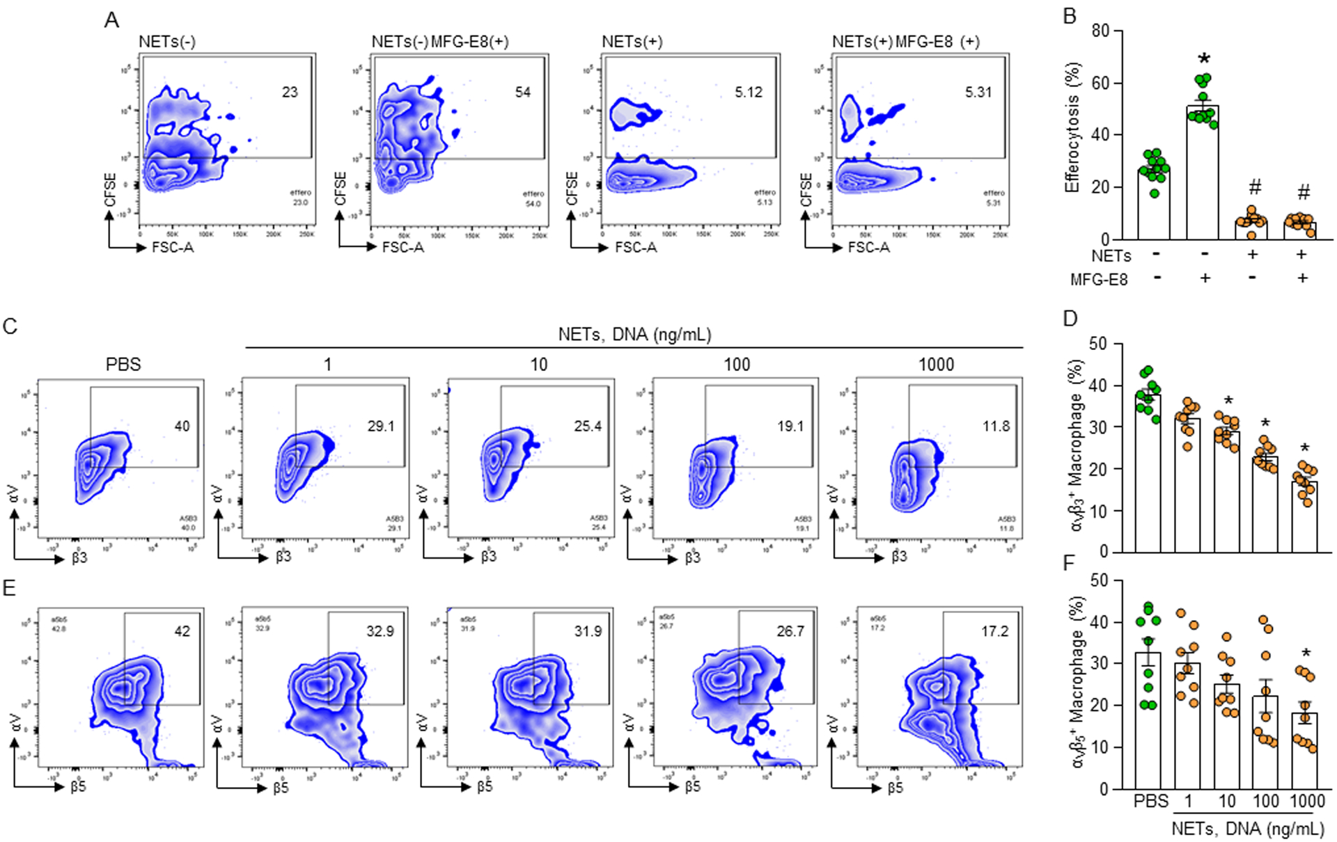
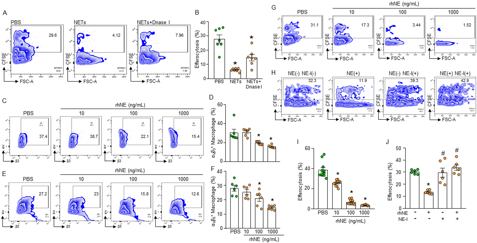
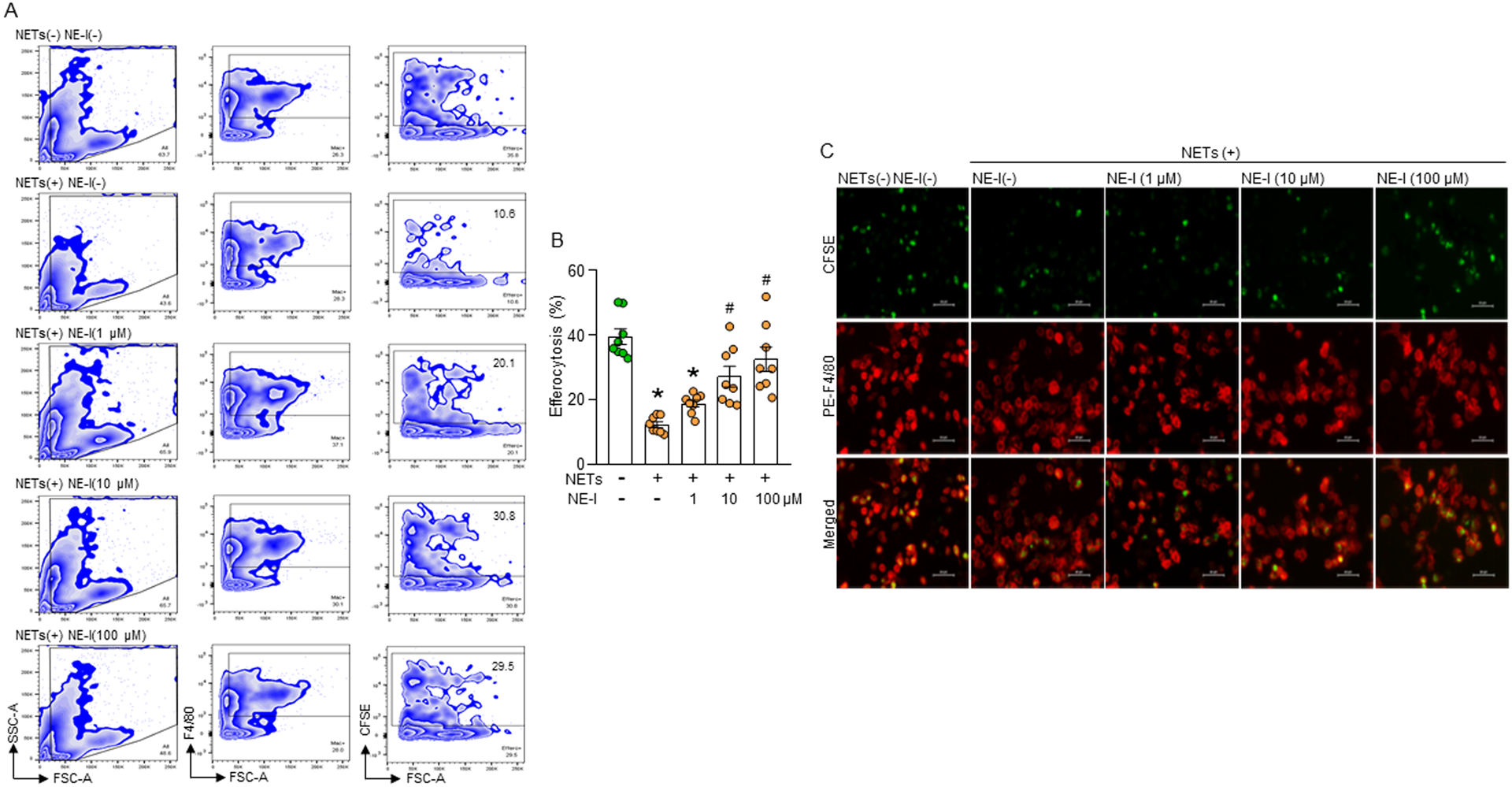
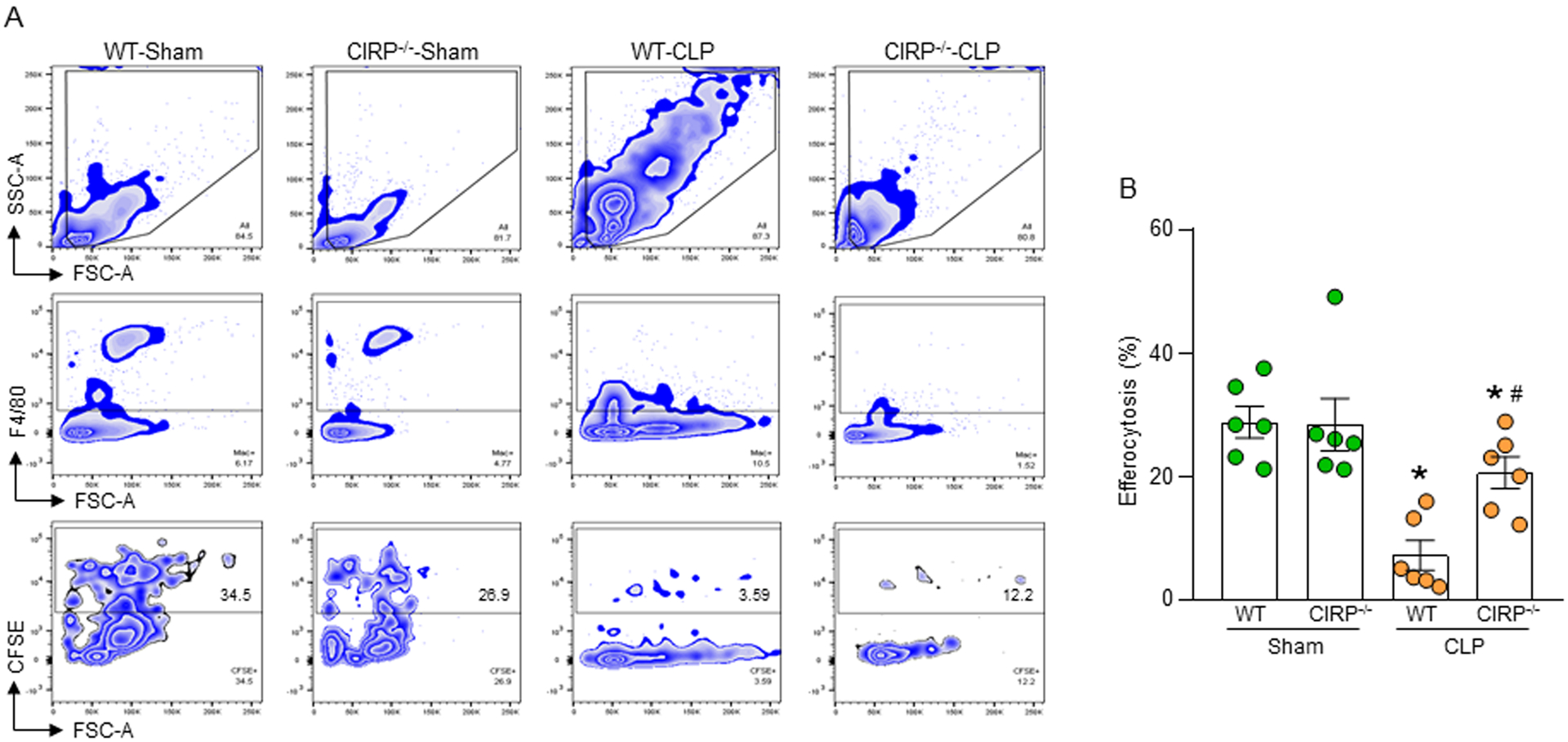
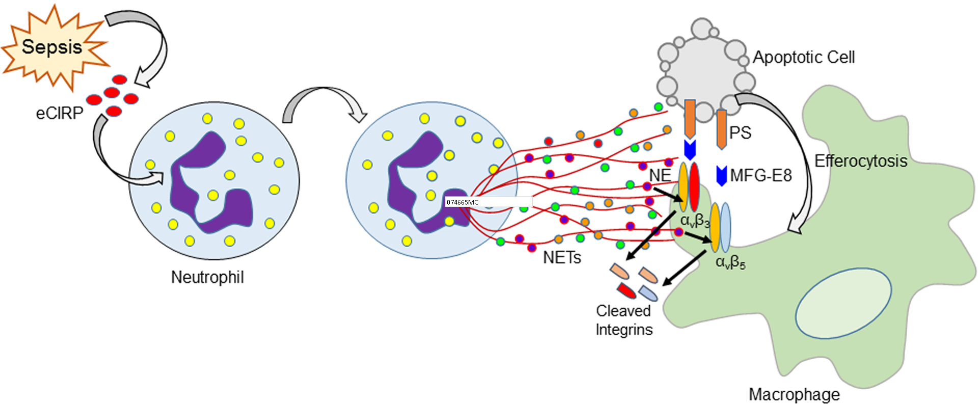
Phagocytic clearance of apoptotic cells by the macrophages (efferocytosis) is impaired in sepsis, but its mechanism is poorly understood. Extracellular cold-inducible RNA-binding protein (eCIRP) is a novel damage-associated molecular pattern that fuels inflammation. We identify that eCIRP-induced neutrophil extracellular traps (NETs) impair efferocytosis through a novel mechanism. Coculture of macrophages and apoptotic thymocytes in the presence of recombinant murine CIRP (rmCIRP)-induced NETs significantly inhibited efferocytosis. Efferocytosis was significantly inhibited in the presence of rmCIRP-treated wild-type (WT), but not PAD4-/- neutrophils. Efferocytosis in the peritoneal cavity of rmCIRP-injected PAD4-/- mice was higher than WT mice. Milk fat globule-EGF-factor VIII (MFG-E8), an opsonin, increased macrophage efferocytosis, whereas the inhibition of efferocytosis by NETs was not rescued upon addition of MFG-E8, indicating disruption of MFG-E8's receptor(s) αvβ3 or αvβ5 integrin by the NETs. We identified neutrophil elastase in the NETs significantly inhibited efferocytosis by cleaving macrophage surface integrins αvβ3 and αvβ5 Using a preclinical model of sepsis, we found that CIRP-/- mice exhibited significantly increased rate of efferocytosis in the peritoneal cavity compared with WT mice. We discovered a novel role of eCIRP-induced NETs to inhibit efferocytosis by the neutrophil elastase-dependent decrease of αvβ3vβ5 integrins in macrophages. Targeting eCIRP ameliorates sepsis by enhancing efferocytosis.


Figures

Figure 1:
Figure 1:. eCIRP-induced NETs inhibit efferocytosis.
(A, B) A total of 5 × 10⁵ peritoneal macrophages were cultured with 1.5 × 10⁶ CFSE-labeled apoptotic cells in presence with PBS or various concentrations of NETs. After 1 h of incubation, cells were washed, fixed with 2% PFA, and efferocytosis was assessed by flow cytometry. Efferocytosis was determined as the percentage of CFSE-positive cells present in F4/80+ macrophages. Data were obtained from 3 independent experiments and expressed as means ± SE (n=7 samples/group). The groups were compared by one-way ANOVA and SNK method (*p<0.05; ***p<0.001 vs. PBS-treated group). (C) Confirmation of phagocytosis of apoptotic cells in F4/80 and CFSE double-positive population by image stream analysis. Representative images showing co-localization of F4/80 and CFSE double positive cells indicate efferocytosis. Scale bar: 7 μm. BMDN, bone marrow-derived neutrophil(s); MPO, myeloperoxidase; CFSE, carboxyfluorescein succinimidyl ester; PFA, paraformaldehyde; NETs, neutrophil extracellular traps; rmCIRP, recombinant mouse cold-inducible RNA-binding protein.
Figure 2:
Figure 2:. eCIRP-induced wild-type, but not PAD4−/− neutrophils inhibit efferocytosis.
(A, B) BMDN (1× 10⁶) isolated from WT or PAD4−/− mice were stimulated with PBS or rmCIRP (1 μg/mL) for 4 h to allow them to form NETs. Peritoneal macrophages (5 × 10⁵) and CFSE-labeled apoptotic cells (Ao) (1.5 × 10⁶) were separately added to PBS- or rmCIRP-treated WT or PAD4−/− PMN. Cells were continued to culture for 1 h. Cells were then stained with PE-F4/80 Abs and assessed efferocytosis by flow cytometry. Data were obtained from 3 independent experiments and expressed as means ± SE (n=7 samples/group). The groups were compared by one-way ANOVA and SNK method. *p<0.05 vs. rmCIRP(−) PMN(−); #p<0.05 vs. rmCIRP(+) WT PMN(+). (C, D) WT and PAD4−/− mice were injected with rmCIRP (5 mg/kg; i.p.). After 4 h of injection with rmCIRP, a total of 1 × 10⁷ CFSE-labeled apoptotic cells were injected i.p. into the mice. After 1 h, peritoneal washout cells were collected, stained with PE-F4/80 Ab, and assessed efferocytosis by flow cytometry. Data were obtained from 3 independent experiments and expressed as means ± SE (n=6 mice/group). The groups were compared by one-way ANOVA and SNK method. *p<0.05 vs. WT mice. (E-J) WT and PAD4−/− mice were injected with rmCIRP (5 mg/kg; i.p.). After 20 h of injecting the mice with rmCIRP, peritoneal washout cells were harvested and stained with macrophage marker PE-F4/80 and neutrophil marker APC-Ly6G Abs. The contents of (E-G) macrophages and (H-J) neutrophils in the peritoneal cavity were assessed my flow cytometry. Data were obtained from 3 independent experiments and expressed as means ± SE (n=6 mice/group). The groups were compared by one-way ANOVA and SNK method. *p<0.05 vs. PBS-treated WT/PAD4−/− mice. PAD4, peptidylarginine deiminase 4; PMN, polymorphonuclear leukocytes; Ao, apoptotic cells.
Figure 3:
Figure 3:. NETs decrease integrin-mediated efferocytosis.
(A, B) Peritoneal macrophages (5 × 10⁵) and CFSE-labeled apoptotic cells (1.5 × 10⁶) in the presence of NETs (1000 ng/mL) with or without rmMFG-E8 (2 μg/mL). After 1 h of incubation, the cells were collected, washed, and stained with PE-F4/80 Ab and assessed efferocytosis in macrophages by flow cytometry. Data were obtained from 3 independent experiments and expressed as means ± SE (n=6 mice/group). The groups were compared by one-way ANOVA and SNK method. *p<0.05 vs. NETs(−) rmMFG-E8(−); #p<0.05 vs. NETs(−) rmMFG-E8(+). (C-F) Impaired surface expression of integrins in NETs-treated macrophages. Peritoneal macrophages (5 × 10⁵) were treated with NETs at various doses. After 1 h of stimulation, the macrophages were washed, stained with (C, D) anti- αvβ3 and (E, F) αvβ5 integrin Abs and detected integrins’ expression by flow cytometry. Data were obtained from 3 independent experiments and expressed as means ± SE (n=9 samples/group). The groups were compared by one-way ANOVA and SNK method. *p<0.05 vs. PBS-treated macrophages. rmMFG-E8, recombinant mouse milk fat globule-EGF-factor VIII.
Figure 4:
Figure 4:. NE cleaves integrins causing impaired efferocytosis.
(A, B) Murine peritoneal macrophages (5 × 10⁵) were cultured with CFSE-stained apoptotic cells (1.5 × 10⁶) in presence of NETs (1000 ng/mL) and DNase I (500U/mL). After 1 h of cell culture, the cells were washed and stained with PE-F4/80 Ab and assessed efferocytosis using flow cytometry. (C-F) Assessment of surface expression of integrins following treatment of the macrophages with various doses of rmNE. After treatment of the peritoneal macrophages (5 × 10⁵) with rmNE for 4 h the cells were washed and stained with (C, D) anti-αvβ3 and (E, F) αvβ5 integrin Abs and assessed their expression by flow cytometry. (G, I) Peritoneal macrophages (5 × 10⁵) and CFSE-labeled apoptotic cells (1.5 × 10⁶) were co-cultured in the presence of various doses of rmNE. After 1 h, the cells were washed and stained with PE-F4/80 Ab and the efferocytosis was assessed by flow cytometry. Data were obtained from 3 independent experiments and expressed as means ± SE (n=7–9 samples/group). The groups were compared by one-way ANOVA and SNK method. *p<0.05 vs. PBS-treated macrophages. (H, J) Peritoneal macrophages (5 × 10⁵) and CFSE-labeled apoptotic cells (1.5 × 10⁶) were co-cultured in the presence of rmNE (1000 ng/mL) with or without NE-I (100 μM). After 1 h, the cells were washed and stained with PE-F4/80 Ab and assessed efferocytosis by flow cytometry. Data were obtained from 3 independent experiments and expressed as means ± SE (n=6 samples/group). The groups were compared by one-way ANOVA and SNK method. *p<0.05 vs. NE(−) NE-I(−); #p<0.05 vs. NE(+) NE-I(−). NE, neutrophil elastase; NE-I, NE-inhibitor.
Figure 5:
Figure 5:. NE inhibitor reverses NET-dependent impairment of efferocytosis.
(A, B) Murine peritoneal macrophages (5 × 10⁵) and CFSE-labeled apoptotic cells (1.5 × 10⁶) in presence of NETs (1000 ng/mL) and various doses of NE-I. After 1 h of cell culture, the cells were washed and stained with PE-F4/80 Ab and assessed efferocytosis using flow cytometry. Data were obtained from 3 independent experiments and expressed as means ± SE (n=8 samples/group). The groups were compared by one-way ANOVA and SNK method. *p<0.05 vs. NETs(−) NE-I(−); #p<0.05 vs. NETs(+) NE-I(–). (C) Murine peritoneal macrophages (5 × 10⁵) were plated in 24-well plate. CFSE-stained apoptotic cells (1.5 × 10⁶) were added to the macrophages. The cells were treated with NETs (1000 ng/mL) and various doses of NE-I. After 1 h of incubation, media was carefully removed, washed the cells with PBS, followed by fixing and stained the cells with PE-F4/80 Ab. Efferocytosis was detected by using fluorescent microscope at ×400 original magnification. Representative images in each group were chosen from 10 randomly captured images. Scale bar: 40 μm.
Figure 6:
Figure 6:. CIRP−/− mice have increased efferocytosis in sepsis.
(A, B) WT and CIRP−/− mice were underwent sham and CLP operation. After 5 h of surgery all mice were injected with CFSE-labeled apoptotic cells (1 × 10⁷) via i.p. injection. After 1 h of injection of apoptotic cells, peritoneal washout cells were collected and stained with PE-F4/80 Ab and assessed efferocytosis by flow cytometry. Data were obtained from 3 independent experiments are expressed as means ± SE (n=6 mice/group). The groups were compared by one-way ANOVA and SNK method. *p<0.05 vs. relevant (WT or CIRP−/−) sham mice; #p<0.05 vs. WT CLP mice.
Figure 7:
Figure 7:. Schematic Summary.
eCIRP is released during sepsis, which induces neutrophils to produce NETs. NE contained in the NETs cleaves αvβ3 and αvβ5 integrins expressed on the surface of macrophages. This leads to decreased binding of apoptotic cell’s opsonin MFG-E8 to the macrophage, thereby impairing the bridge formation between apoptotic cell and macrophage and inhibiting efferocytosis.

References

    1. Singer M, Deutschman CS, Seymour CW, Shankar-Hari M, Annane D, Bauer M, Bellomo R, Bernard GR, Chiche JD, Coopersmith CM, Hotchkiss RS, Levy MM, Marshall JC, Martin GS, Opal SM, Rubenfeld GD, van der Poll T, Vincent JL, and Angus DC. 2016. The Third International Consensus Definitions for Sepsis and Septic Shock (Sepsis-3). JAMA 315: 801–810. - PMC - PubMed
    1. Aziz M, Jacob A, Yang WL, Matsuda A, and Wang P. 2013. Current trends in inflammatory and immunomodulatory mediators in sepsis. J Leukoc Biol 93: 329–342. - PMC - PubMed
    1. Wesche DE, Lomas-Neira JL, Perl M, Chung CS, and Ayala A. 2005. Leukocyte apoptosis and its significance in sepsis and shock. J Leukoc Biol 78: 325–337. - PubMed
    1. Elliott MR, Koster KM, and Murphy PS. 2017. Efferocytosis Signaling in the Regulation of Macrophage Inflammatory Responses. J Immunol 198: 1387–1394. - PMC - PubMed
    1. Proto JD, Doran AC, Gusarova G, Yurdagul A, Sozen E, Subramanian M, Islam MN, Rymond CC, Du J, Hook J, Kuriakose G, Bhattacharya J, and Tabas I. 2018. Regulatory T Cells Promote Macrophage Efferocytosis during Inflammation Resolution. Immunity 49: 666–677.e666. - PMC - PubMed

Publication types