Review
. 2021 Feb;32(2):268-278.
doi: 10.1681/ASN.2020071082. Epub 2020 Dec 30.

New 'Antigens' in Membranous Nephropathy

Affiliations
Review

New 'Antigens' in Membranous Nephropathy

Sanjeev Sethi. J Am Soc Nephrol. 2021 Feb.

Abstract

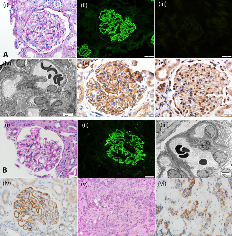
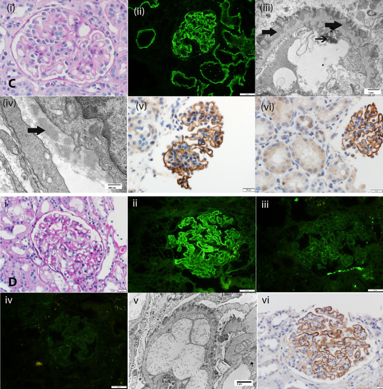
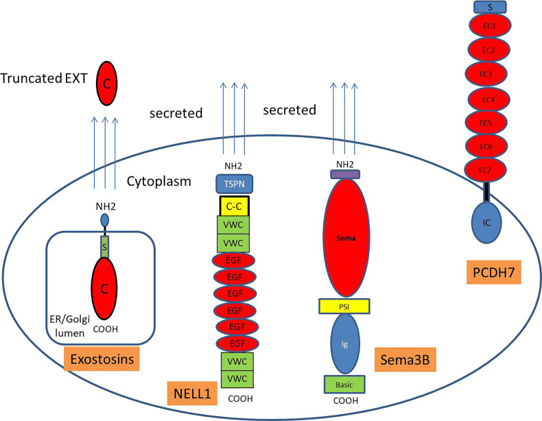
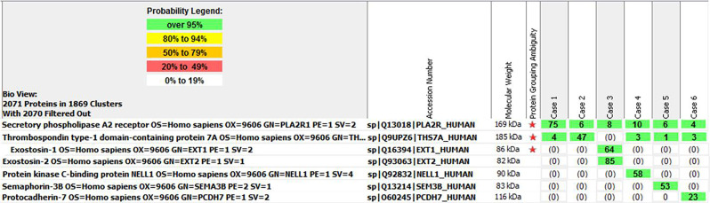
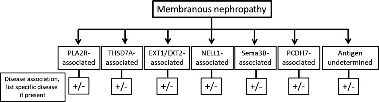
Membranous nephropathy (MN) occurs due to deposition of immune complexes along the subepithelial region of glomerular basement membrane. Two previously identified target antigens for the immune complexes, PLA2R (identified in 2009) and THSD7A (in 2014), account for approximately 60% of all MN, both primary and secondary. In the remaining MN, target antigens were unknown. Use of laser microdissection and mass spectrometry enabled identification of new "antigens." This approach led to the identification of four novel types of MN: exotosin 1 (EXT1)- and exotosin 2 (EXT2)-associated MN, NELL1-associated MN, Sema3B-associated MN, and PCDH7-associated MN. Each of these represents a distinct disease entity, with different clinical and pathologic findings. In this review, the structure of the proteins and the clinical and pathologic findings of the new types of MN are discussed. The role of mass spectrometry for accurate diagnosis of MN cannot be overemphasized. Finally, any classification of MN should be made on the basis of the antigens that are detected. Further studies are required to understand the pathophysiology, response to treatment, and outcomes of these new MNs.

Keywords: kidney biopsy; membranous nephropathy; nephrotic syndrome.


Figures

Figure 1.
Figure 1.
Biopsy specimen findings of EXT1/EXT2-, NELL1-, Sema3B-, and PCDH7-associated MN. (A) EXT1/EXT2-associated MN: (i) Periodic acid–Schiff stain showing thickened glomerular capillary walls. Original magnification, ×40. (ii) IF microscopy showing bright C3 staining along glomerular capillary walls; (iii) IF microscopy is negative for PLA2R; (iv) electron microscopy showing subepithelial electron dense deposits. Original magnification, ×2900. IHC showing (v) EXT1 and (vi) EXT2 staining along the GBM. (B) NELL1-associated MN: (i) Periodic acid–Schiff stain showing thickened glomerular capillary walls. Original magnification, ×40. (ii) IF microscopy showing bright C3 staining along glomerular capillary walls; (iii) electron microscopy showing subepithelial electron dense deposits. Original magnification, ×2900. (iv) IHC showing NELL1 staining along the glomerular capillary walls; (v) hematoxylin and eosin stain showing squamous cell carcinoma; (vi) IHC showing that the tumor cells are positive for NELL1. (C) Sema3B-associated MN: (i) Periodic acid–Schiff stain showing thickened glomerular capillary walls. Original magnification, ×40. (ii) IF microscopy showing bright IgG staining along the GBM and TBM. Original magnification, ×20. (iii) Electron microscopy showing subepithelial electron dense deposits along GBM and tubuloreticular inclusions in endothelial cells. Original magnification, ×11,000. (iv) Electron microscopy showing TBM electron dense deposits. Original magnification, ×30,000. (v) IHC showing Sema3B staining along the GBM. Original magnification, ×40. (vi) IHC showing Sema3B staining along GBM but negative staining along the TBM. Original magnification, ×20. Arrows point to tubular basement membrane deposits. (D) PCDH7-associated MN: (i) Periodic acid–Schiff stain showing thickened glomerular capillary walls. Original magnification, ×40. (ii) IF microscopy showing bright IgG staining along the GBM. Original magnification, ×240. (iii) IF microscopy showing trace staining for C3, and (iv) negative staining for C1q. (v) Electron microscopy showing subepithelial electron dense deposits alongGBM. Original magnification, ×6800. (vi) IHC showing PCDH7 staining along the GBM. Original magnification, ×40.
Figure 1.
Figure 1.
Biopsy specimen findings of EXT1/EXT2-, NELL1-, Sema3B-, and PCDH7-associated MN. (A) EXT1/EXT2-associated MN: (i) Periodic acid–Schiff stain showing thickened glomerular capillary walls. Original magnification, ×40. (ii) IF microscopy showing bright C3 staining along glomerular capillary walls; (iii) IF microscopy is negative for PLA2R; (iv) electron microscopy showing subepithelial electron dense deposits. Original magnification, ×2900. IHC showing (v) EXT1 and (vi) EXT2 staining along the GBM. (B) NELL1-associated MN: (i) Periodic acid–Schiff stain showing thickened glomerular capillary walls. Original magnification, ×40. (ii) IF microscopy showing bright C3 staining along glomerular capillary walls; (iii) electron microscopy showing subepithelial electron dense deposits. Original magnification, ×2900. (iv) IHC showing NELL1 staining along the glomerular capillary walls; (v) hematoxylin and eosin stain showing squamous cell carcinoma; (vi) IHC showing that the tumor cells are positive for NELL1. (C) Sema3B-associated MN: (i) Periodic acid–Schiff stain showing thickened glomerular capillary walls. Original magnification, ×40. (ii) IF microscopy showing bright IgG staining along the GBM and TBM. Original magnification, ×20. (iii) Electron microscopy showing subepithelial electron dense deposits along GBM and tubuloreticular inclusions in endothelial cells. Original magnification, ×11,000. (iv) Electron microscopy showing TBM electron dense deposits. Original magnification, ×30,000. (v) IHC showing Sema3B staining along the GBM. Original magnification, ×40. (vi) IHC showing Sema3B staining along GBM but negative staining along the TBM. Original magnification, ×20. Arrows point to tubular basement membrane deposits. (D) PCDH7-associated MN: (i) Periodic acid–Schiff stain showing thickened glomerular capillary walls. Original magnification, ×40. (ii) IF microscopy showing bright IgG staining along the GBM. Original magnification, ×240. (iii) IF microscopy showing trace staining for C3, and (iv) negative staining for C1q. (v) Electron microscopy showing subepithelial electron dense deposits alongGBM. Original magnification, ×6800. (vi) IHC showing PCDH7 staining along the GBM. Original magnification, ×40.
Figure 2.
Figure 2.
Schematic representation of the MN proteins. Exostosins are transmembrane proteins in the Golgi apparatus that have a short amino-terminal cytoplasmic tail (NH2), a single transmembrane domain, a stem/stalk region (S), and a long globular catalytic C-terminal domain (C) within the Golgi lumen. Exostosins are secreted as truncated (C) proteins. NELL1 is a secreted protein and is characterized by an amino-terminal TSP-1-like (TSPN) domain, a coiled-coil (C-C) domain, two von Willebrand factor type C (VWC) domains, six EGF-like domains (E), and two VWC domains. Sema3B is a secreted protein with large sema domain region, plexin-semaphorin-integrin (PSI) domain, Ig domain, and short C-terminal basic domain. PCDH7 is a transmembrane protein with a signal (S) peptide, seven extracellular (EC) domains, a single pass transmembrane domain (black bar), and an intracellular (IC) cytoplasmic domain. COOH, carboxyl group; ER, endoplasmic reticulum.
Figure 3.
Figure 3.
Cumulative incidence of ESKD in patients with lupus membranous nephritis (LMN). Kaplan–Meier plots of the cumulative incidence of ESKD over 10 years. (A) EXT1/EXT2-positive and EXT1/EXT2-negative LMN (including class III/IV lupus nephritis [LN]): 64 EXT+ versus 96 EXT−; two versus 18 events; time to event, 116 versus 101 months; P=0.007. (B) EXT1/EXT2-positive and EXT1/EXT2-negative pure class V LMN (with no class III/IV LN): 48 EXT+ versus 65 EXT−; two versus 11 events; time to event, 115 versus 104 months; P=0.08. (C) EXT1/EXT2-positive and EXT1/EXT2-negative class V LMN + class III/IV LN: 16 EXT+ versus 31 EXT−; zero versus seven events; time to event in EXT−, 96 months; P=0.03. EXT1/EXT-positive represented by dotted lines and EXT1/EXT2-negative represented by solid lines. Plots courtesy of Dr. Aishwarya Ravindran and Dr. Marta C. Moura.
Figure 4.
Figure 4.
Representative mass spectrometry and detection of antigens in MN. Case 1 is from a biopsy specimen of PLA2R-associated MN, case 2 from THSD7A-associated MN, case 3 from EXT1/EXT2-associated MN, case 4 from NELL1-associated MN, case 5 from Sema3B-associated MN, and case 6 from PCDH7-associated MN. Numbers in green boxes represent spectral counts of MS/MS matches to a respective protein. Red star indicates shared amino acid sequences among proteins.
Figure 5.
Figure 5.
Proposed classification of MN. MN is classified on the basis of the antigen detected. In cases where none of the known antigens are detected, the terminology “undetermined” should be used. The disease association should be given if present, not present, or if not known.

References

    1. Heymann W, Hackel DB, Harwood S, Wilson SG, Hunter JL: Production of nephrotic syndrome in rats by Freund’s adjuvants and rat kidney suspensions. Proc Soc Exp Biol Med 100: 660–664, 1959 - PubMed
    1. Beck LH Jr., Bonegio RGB, Lambeau G, Beck DM, Powell DW, Cummins TD, et al. .: M-type phospholipase A2 receptor as target antigen in idiopathic membranous nephropathy. N Engl J Med 361: 11–21, 2009 - PMC - PubMed
    1. Tomas NM, Beck LH Jr., Meyer-Schwesinger C, Seitz-Polski B, Ma H, Zahner G, et al. .: Thrombospondin type-1 domain-containing 7A in idiopathic membranous nephropathy. N Engl J Med 371: 2277–2287, 2014 - PMC - PubMed
    1. Couser WG: Primary membranous nephropathy. Clin J Am Soc Nephrol 12: 983–997, 2017 - PMC - PubMed
    1. Sethi S, Theis JD: Pathology and diagnosis of renal non-AL amyloidosis. J Nephrol 31: 343–350, 2018 - PubMed

MeSH terms

LinkOut - more resources