. 2020 Dec 7;2020(45):7087-7100.
doi: 10.1002/ejoc.202001211. Epub 2020 Nov 16.

Double Porphyrin Cage Compounds

Affiliations

Double Porphyrin Cage Compounds

Kathleen Stout et al. European J Org Chem. .

Abstract

The synthesis and characterization of double porphyrin cage compounds are described. They consist of two porphyrins that are each attached to a diphenylglycoluril-based clip molecule via four ethyleneoxy spacers, and are linked together by a single alkyl chain using "click"-chemistry. Following a newly developed multistep synthesis procedure we report three of these double porphyrin cages, linked by spacers of different lengths, i.e. 3, 5, and 11 carbon atoms. The structures of the double porphyrin cages were fully characterized by NMR, which revealed that they consist of mixtures of two diastereoisomers. Their zinc derivatives are capable of forming sandwich-like complexes with the ditopic ligand 1,4-diazabicyclo[2,2,2]octane (dabco).

Keywords: Axial ligands; Host–guest chemistry; Porphyrin compounds.


Figures

Figure 1
Figure 1
(A) Molecular structure of catalytic porphyrin cage compound MnSC and the axial ligand bupy. (B) Schematic representation of the processive catalytic conversion of cis‐polybutadiene (yellow) into its polyepoxide, carried out by MnSC (blue), to which a bupy ligand (green) is axially coordinated. (C) Schematic representation of a double porphyrin cage compound (yellow) in which the “writing” cage serves as a processive catalyst for the epoxidation of a polymer chain (red) and the “instructing” cage binds a Me2V cofactor (blue), which transfers information via the ditopic ligand dabco (orange) to the “writing cage”. (D) Molecular structures of the double porphyrin cage compounds described in this paper.
Scheme 1
Scheme 1
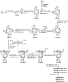
Synthesis of double porphyrin cage compounds.
Figure 2
Figure 2
(A) 1H NMR spectrum of H4C3DC in CDCl3 and proton/carbon numbering of the compound. The Roman numbers I, II, III and IV and the related colors indicate the four symmetry quandrants within the porphyrin cage structure. (B) 15N spectrum of H4C3DC in CDCl3.
Figure 3
Figure 3
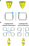
(A) Representations of the two enantiomers of monofunctionalized single cage compounds 8–11. Top: 3D representations. Bottom: schematic view of the porphyrin (black square) seen from the top. The black lines left and right from the square indicate the location of the o‐xylylene side walls of the cage, the cyan dot the functional group at the top. (B) Representations (top views and 3D views) of the two diastereoisomers that are obtained by the coupling of the enantiomers of monofunctionalized single cage compound 11.
Figure 4
Figure 4
(A) Changes in UV/Vis spectra of Zn2C3DC upon the addition of dabco, in CHCl3/CH3CN, 1:1 (v/v). (B) Titration curves extracted from the spectral changes in (A), monitoring the absorbance of the bands at 426 (red) and 431 nm (blue). The solid lines through the data points represent fits assuming a standard 1:2 binding model.
Figure 5
Figure 5
Computer‐modeled structures of the sandwich complexes of double zinc porphyrin cages (indigo) (A) Zn2C3DC, (B) Zn2C5DC and (C) Zn2C11DC with dabco (red). The alkyl spacers between the cages are indicated in yellow.
Figure 6
Figure 6
1H NMR titration of Zn2C3DC with 0 to 8 equiv. of dabco (500 MHz, 298 K, CDCl3/CD3CN, 1:1 (v/v)). The blue box and arrow indicate the location of the signal of dabco in the 1:1 sandwich complex, and the red arrow the signal of non‐coordinated dabco.
Figure 7
Figure 7
Variable temperature 1H NMR spectra of the 1:1 sandwich complex between Zn2C3DC and dabco (400 MHz, CDCl3/CD3CN, 1:1 (v/v)). The arrow indicates the location of the signal of dabco in the 1:1 sandwich complex.
Figure 8
Figure 8
1H NMR titration of Zn2C3DC with 0 to 4 equiv. of dabco (500 MHz, 248 K, CDCl3/CD3CN, 1:1 (v/v)). The blue and purple arrows indicate the locations of the signals of dabco in the 1:1 sandwich complex (–4.58 ppm) and in the 2:1 open‐folded complex (–2.92 ppm), respectively.
Figure 9
Figure 9
Carbon, proton, and nitrogen numbering of 5‐(5‐bromo‐2‐methoxyphenyl)‐10,15,20‐tris(2‐methoxyphenyl) porphyrin 5 (left) and the double cage compounds M2CnDC (M = 2H or Zn, n = 3, 5 or 11) (right) used for all NMR analyses. For the double cage compounds, R represents the mirror image of the cage molecule, with the restriction that for Zn2C3DC the attachment starts at carbon number 63, for Zn2C5DC at carbon atom 64, and for Zn2C11DC at carbon atom 67 (the latter is shown). The CH2‐groups of the diphenylglycoluril framework (carbon atoms 45, 46, 48 and 50) all have two inequivalent geminal protons, marked as a and b. The location of the signals of these protons could only be identified for CH2‐50, with the help of ROESY experiments.

References

    1. Johnson A. and O'Donnell M., Annu. Rev. Biochem, 2005, 74, 283–315. - PubMed
    1. a) van Dongen S. F. M., Elemans J. A. A. W., Rowan A. E. and Nolte R. J. M., Angew. Chem. Int. Ed, 2014, 53, 11420–11428; - PubMed
    2. Angew. Chem, 2014, 126, 11604;
    3. b) Kovall R. and Matthews B. W., Science, 1997, 277, 1824–1827. - PubMed
    1. a) Elemans J. A. A. W., Bijsterveld E. J. A., Rowan A. E. and Nolte R. J. M., Chem. Commun, 2000, 2443–2444;
    2. b) Elemans J. A. A. W., Bijsterveld E. J. A., Rowan A. E. and Nolte R. J. M., Eur. J. Org. Chem, 2007, 751–757.
    1. a) Thordarson P., Bijsterveld E. J. A., Rowan A. E. and Nolte R. J. M., Nature, 2003, 424, 915–918; - PubMed
    2. b) Bernar I., Rutjes F. P. J. T., Elemans J. A. A. W. and Nolte R. J. M., Catalysts, 2019, 9, 195;
    3. c) Monnereau C., Hidalgo Ramos P., Deutman A. B. C., Elemans J. A. A. W., Nolte R. J. M. and Rowan A. E., J. Am. Chem. Soc, 2010, 132, 1529–1531. - PubMed
    1. a) Martens S., Landuyt A., Espeel P., Devreese B., Dawyndt P. and Du Prez F., Nat. Commun, 2018, 9, 4451; - PMC - PubMed
    2. b) Szweda R., Tschopp M., Felix O., Decher G. and Lutz J.‐F., Angew. Chem. Int. Ed, 2018, 57, 15817–15821; - PubMed
    3. Angew. Chem, 2018, 130, 16043;
    4. c) Rutten M. G. T. A., Vaandrager F. W., Elemans J. A. A. W. and Nolte R. J. M., Nat. Rev. Chem, 2018, 2, 365–381.

LinkOut - more resources