Review
. 2020 Dec 16;12(24):6082-6102.
doi: 10.1002/cctc.202001107. Epub 2020 Oct 1.

Looking Back: A Short History of the Discovery of Enzymes and How They Became Powerful Chemical Tools

Affiliations
Review

Looking Back: A Short History of the Discovery of Enzymes and How They Became Powerful Chemical Tools

Christian M Heckmann et al. ChemCatChem. .

Abstract

Enzymatic approaches to challenges in chemical synthesis are increasingly popular and very attractive to industry given their green nature and high efficiency compared to traditional methods. In this historical review we highlight the developments across several fields that were necessary to create the modern field of biocatalysis, with enzyme engineering and directed evolution at its core. We exemplify the modular, incremental, and highly unpredictable nature of scientific discovery, driven by curiosity, and showcase the resulting examples of cutting-edge enzymatic applications in industry.

Keywords: enzymes; green chemistry; history of biocatalysis; immobilization; protein engineering.


Conflict of interest statement

The authors declare no conflict of interest.

Figures

Figure 1
Figure 1
Timeline of major developments in enzymology, molecular biology, and biocatalysis.
Scheme 1
Scheme 1
Synthesis of L‐ephedrine: Benzoin‐type addition of acetaldehyde (formed in‐situ by yeast metabolism) to benzaldehyde (now known to be catalyzed by pyruvate decarboxylase), [14] followed by reductive amination with methylamine. [11]
Figure 2
Figure 2
Plots of the Michaelis‐Menten model, illustrating equation 1. A) Velocity vs. substrate concentration under constant enzyme concentration. The substrate affinity Km corresponds to the substrate concentration at which the reaction reaches half of vmax, which is the velocity at infinite substrate concentration. B) vmax vs. enzyme concentration. The reaction is first‐order with respect to enzyme concentration, with the rate constant kcat.
Figure 3
Figure 3
The original drawings of an α‐helix (left) and parallel and anti‐parallel β‐sheets (right), published by Pauling in 1951.[ 24b , 24d ]
Figure 4
Figure 4
Top left: Clay model of the first X‐ray structure of a protein, myoglobin, at 6 Å resolution. [26a] Right: electron density sections of myoglobin at 2 Å resolution and sketch of groups coordinated to iron. [26b] Bottom left: Model of the catalytic triad and oxyanion hole of chymotrypsin, as inferred from crystal‐structures. [31c]
Figure 5
Figure 5
Selected structures of common cofactors that were known by 1955: NAD+, NADP+ and FAD are redox catalysts, ATP transfers energy released during glycolysis, thiamine is the co‐factor of pyruvate decarboxylase during fermentation, and pyridoxal is the co‐factor of transaminases, which are of particular industrial importance, as well as racemases, decarboxylases, and lysases involved in amino acid metabolism.
Figure 6
Figure 6
Selected mechanisms of co‐factors that were being elucidated in the 1950s: enantiospecificity during hydride transfer from NAD(P)H in alcohol dehydrogenases, and thiamine‐dependent decarboxylation. [33b]
Figure 7
Figure 7
Mechanism of transamination. For clarity, the individual steps of aldimine/ketimine formation and hydrolysis as well as transimination are not shown. The mechanism is symmetric – referred to as a “ping‐pong” bi‐bi or shuttle mechanism – and fully reversible. Note: the ketimine intermediates are a second aldimine if one of the R‐groups is a hydrogen. Catalytic lysine: red; amine donor: blue; ketone acceptor: green.
Scheme 2
Scheme 2
Top: Isomerization of d‐glucose to d‐fructose, catalyzed by an immobilized glucose isomerase as used in the production of HFCS. Bottom: Hydrolysis of Penicillin to give 6‐APA, which can then be acylated to give several semi‐synthetic antibiotics.
Figure 8
Figure 8
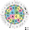
The genetic code. Codons consisting of three bases (triplets) correspond to different amino acids. Each amino acid may be spelled by multiple codons (the code is degenerate). The chart is read from the inside outwards (following the red arrows), e. g. “AUG” corresponds to the start codon, methionine, corresponding to the beginning of a protein. The genetic code is universal, i. e. identical across all organisms with only a few exceptions. https://commons.wikimedia.org/wiki/File:Aminoacids_table.svg, public domain.
Figure 9
Figure 9
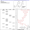
Principle of Sanger sequencing: A DNA strand (blue) is copied by DNA polymerase. If a small quantity of dideoxynucleotides (ddNTP) is offered in addition to deoxynucleotides (for example ddCTP), chains will terminated whenever a ddCTP is incorporated instead of a dCTP, as the 3′‐OH needed for chain‐extension is missing. If this experiment is repeated for all four nucleotides, and the products are separated by size, the sequence of the DNA template can be inferred. Modern Sanger sequencing includes all four ddNTPs in a single sequencing reaction, and distinguishes incorporation of the different bases at the termination site via fluorescent labels, such as the label (red) for the ddT – BigDye terminator shown. [60]
Figure 10
Figure 10
Type I restriction enzymes cut at a non‐defined remote location from the recognition site (example of EcoKI). Type II restriction enzymes cut at a well‐defined position within (or close to) the recognition sequence, often in a staggered way, producing cohesive ends (example of HindIII).
Figure 11
Figure 11
Left: Map of pBR322, showing the unique restriction sites inside both antibiotic resistance genes. Right: Generic map of a typical (empty) expression vector, having an origin of replication (for replication in vivo), a selective marker (Ampicillin resistance in this instance), and the T7 promoter and terminator flanking a His‐tag (to allow purification of the insert) and the multiple cloning site, which contains a large number of unique restriction sites (not shown) for easy cloning.
Scheme 3
Scheme 3
Application of penicillin acylase for the synthesis of amoxicillin and ampicillin from 6‐APA. X=NH2 or OMe.
Figure 12
Figure 12
The principles of PCR: DNA is denatured at high temperature, primers supplied in the reaction mixture are annealed, and the template is copied. Repeated cycles exponentially amplify the target sequence. Site‐directed mutagenesis: a mutation is incorporated in the primer; the amplified product now contains the changed base‐pair. epPCR: a polymerase that occasionally incorporates incorrect nucleotides is used. The product now contains a set of different sequences that differ from the parent in a few positions. Recombination: several sequences are shuffled to produce a diverse set of new sequences from the parents.
Figure 13
Figure 13
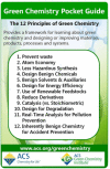
The 12 principles of green chemistry, reproduced with permission from ACS Green Chemistry Institute® (https://www.acs.org/content/acs/en/greenchemistry/principles/12‐principles‐of‐green‐chemistry.html). Copyright 2020 American Chemical Society.
Scheme 4
Scheme 4
The lipase‐catalyzed BASF process for the kinetic resolution of amines. Enantioselectivity is often essentially perfect and conversions quantitative, the amide and amine can be separated by distillation, the amide is readily hydrolyzed (giving access to both enantiomers), and the undesired enantiomer can be racemized and recycled. The process can also be run neat (e. g. in the case of 1‐methoxy‐2‐aminopropane). [108b] Other esters than the ethyl may be used; however, the methoxy‐group is critical for an efficient reaction.
Figure 14
Figure 14
Evolution of ATA‐117 to produce sitagliptin, [123] compared to the chemocatalytic route using a rhodium catalyst. Over 11 rounds of evolution, the conditions of the screening (substrate loading, temperature, cosolvent concentration (Rd 3–6 MeOH, otherwise DMSO) were gradually increased to the process level. Overlaid is the steady increase in conversion under process conditions, as well as the increase in the total number of mutations (note, several mutations changed throughout the process).
Figure 15
Figure 15
Engineering of an IRED for the synthesis of GSK2879552, and the alternative chemical route. Insert: improvement of the catalyst over 3 rounds of evolution; acceptable operating space (black dotted line), wild type IR‐46 (grey), M1 (orange), M2 (green), M3 at small scale (blue) and process scale (red). Adapted from Schober et al. [124]
Figure 16
Figure 16
Top: Exploiting enzyme promiscuity to evolve new catalytic activity. Enzymes often exhibit promiscuous activity toward non‐native substrates or reactions. By applying directed evolution, a “specialist” enzyme might be transformed into another specialist enzyme for the new activity, at the cost of diminishing its original function. Such a transformation proceeds through a “low‐fitness valley” where the enzyme is not very good at either the new or the original function. Figure reproduced from ref. [93] Bottom: This concept was applied to evolve a cytochrome c from Rhodothermus marinus (without any native catalytic function) to catalyze Si−H carbene insertions. [126] The final variant was 33x more active than the parent and became more specialized for Si−H insertion over N−H insertion chemistry, both promiscuous activities of the wt.
Figure 17
Figure 17
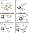
Examples of enzyme immobilization strategies. A) Mechanical entrapment restricts the diffusion of the enzyme. B) Adsorption through ionic interactions, offering little control over the orientation of the enzyme. C) Adsorption though affinity, in this case His‐tag‐metal coordination, allowing control of over the orientation of the enzyme through the tag placement. D) Covalent attachment, offering little control over the orientation of the enzyme. Multipoint attachment can lead to irreversible deformation of the enzyme shape. Common functional groups for covalent attachment are carboxylic acids, aldehydes, and epoxides – using amide formation, reductive amination, and ring opening, respectively. E) Affinity‐directed covalent immobilization orients the enzyme prior to covalent attachment. F) By fusing a (small) protein to the enzyme, covalent immobilization and any shape disruption can be localized to that fusion protein, reducing the effect on the enzyme. However, such an enzyme is more exposed to the environment and stability benefits from immobilization may be diminished. Not shown are covalent crosslinking of enzymes (e. g. using a dialdehyde), and the inherent different properties of supports, with respect to e. g. their size, pore‐size, hydrophilicity/hydrophobicity, etc.
Scheme 5
Scheme 5
Competing acyl transfer (red) and hydrolysis reactions (blue) catalyzed by Penicillin acylase. By tuning the characteristics of the support, synthesis can be kinetically favored over the thermodynamically favored hydrolysis reaction.
Scheme 6
Scheme 6
Three examples of enzymatic cascades. A) Transformation of amines into alcohols, using an immobilized transaminase and either an ADH or KRED in flow. [142] B) A Suzuki cross‐coupling to produce a bi‐aryl ketone which is then aminated using a transaminase catalyst. The transaminase had to tolerate 30 % DMF carried over from the cross‐coupling, as well Pd catalyst, excess base, and unreacted boronic acid. [143] C) halogenation of aromatic compounds using a halogenase, followed by a Suzuki coupling. The enzyme had to be separated, either by ultrafiltration, immobilization, or compartmentalization from the Pd catalyst. [144]
Figure 18
Figure 18
Nine‐enzyme cascade to produce the HIV drug islatravir. Five enzymes had to be evolved. Compared to a chemical synthesis, steps were reduced by more than half and yield was almost doubled. No purification of intermediates was necessary. Immobilized enzymes shown attached to spheres. Figure adapted from Huffman et al. [145]

References

    1. McGovern P. E., Zhang J., Tang J., Zhang Z., Hall G. R., Moreau R. A., Nuñez A., Butrym E. D., Richards M. P., Wang C. S., Cheng G., Zhao Z., Wang C., Proc. Natl. Acad. Sci. USA 2004, 101, 17593–17598. - PMC - PubMed
    1. Armstrong E. F., Nature 1933, 131, 535–537.
    1. E. Büchner, “Eduard Büchner – Nobel Lecture: Cell-free fermentation,” can be found under https://www.nobelprize.org/prizes/chemistry/1907/buchner/lecture/, 1907.
    1. W. Kühne, in Verhandlungen Des Naturhistorisch-Medicinischen Vereins Zu Heidelb, 1877, pp. 190–193.
    1. Robertson A. J. B., Platinum Met. Rev. 1975, 19, 64–69.

LinkOut - more resources