

A Solitary Choroidal Mass with Spontaneous Resolution
- PMID: 33381337
- PMCID: PMC7748891
- DOI: 10.1155/2020/8882617
A Solitary Choroidal Mass with Spontaneous Resolution
Abstract
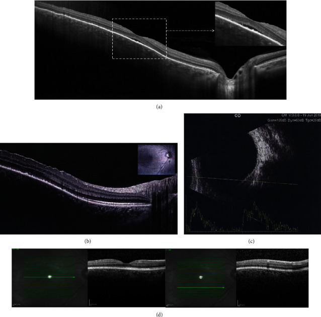
Background: To report an atypical case of a transient choroidal mass lesion with spontaneous resolution. Case Presentation. A solitary choroidal mass with an overlying neurosensory retinal detachment was seen in an otherwise healthy 31-year-old female. General physical examinations and serum chemistry were unremarkable. The patient had spontaneous resolution two weeks after initial examination without treatment.
Conclusions: Inflammatory choroidal masses may be self-limited, but complete diagnostic measures must always be performed in these patients to distinguish between important causes such as tuberculosis, sarcoidosis, and tumors.
Copyright © 2020 Fariba Ghassemi et al.
Conflict of interest statement
None of the authors have any conflicts of interest related to this study.
Figures

Similar articles
-
Solitary choroidal mass as the presenting sign in systemic sarcoidosis.Br J Ophthalmol. 1983 Dec;67(12):826-9. doi: 10.1136/bjo.67.12.826. Br J Ophthalmol. 1983. PMID: 6671099 Free PMC article.
-
Ocular sarcoidosis presenting as a solitary choroidal mass.Can J Ophthalmol. 1992 Feb;27(1):25-9. Can J Ophthalmol. 1992. PMID: 1555132 Review.
-
Two Patients with Atypical Choroidal Detachment.Case Rep Ophthalmol. 2021 May 3;12(2):315-319. doi: 10.1159/000513220. eCollection 2021 May-Aug. Case Rep Ophthalmol. 2021. PMID: 34054477 Free PMC article.
-
DIAGNOSTIC CHALLENGES IN NECROTIC UVEAL MELANOMA.Retin Cases Brief Rep. 2022 Jan 1;16(1):92-94. doi: 10.1097/ICB.0000000000000913. Retin Cases Brief Rep. 2022. PMID: 31425450
-
[Choroidal granuloma in sarcoidosis. Discussion apropos of 2 cases].J Fr Ophtalmol. 1988;11(11):773-8. J Fr Ophtalmol. 1988. PMID: 3074978 Review. French.
References
Publication types
LinkOut - more resources
Full Text Sources
