Review
. 2020 Dec;10(6):1918-1945.
doi: 10.21037/cdt-20-530.

Cardiac CT angiography: normal and pathological anatomical features-a narrative review

Affiliations
Review

Cardiac CT angiography: normal and pathological anatomical features-a narrative review

Alberto Clemente et al. Cardiovasc Diagn Ther. 2020 Dec.

Abstract

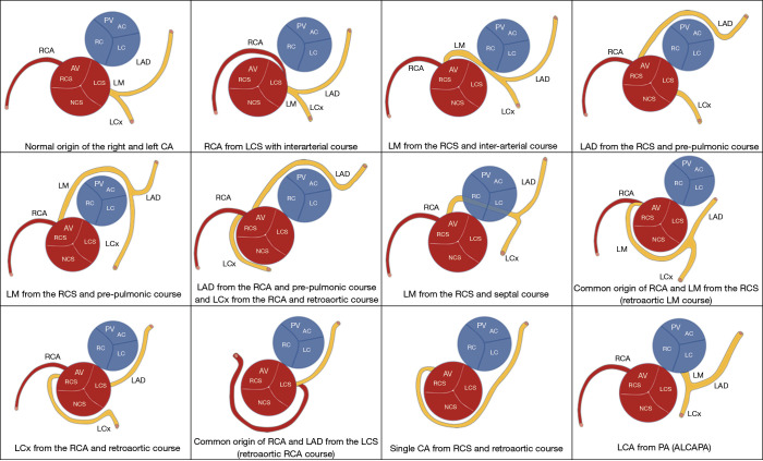
The normal and pathological anatomy of the heart and coronary arteries are nowadays widely developed topics and constitute a fundamental part of the cultural background of the radiologist. The introduction of cardiac ECG-gated synchronized CT scanners with an ever-increasing number of detectors and with increasingly high structural characteristics (increase in temporal resolution, increase in contrast resolution with dual-source, dual energy scanners) allows the virtual measurement of anatomical in vivo structures complying with heart rate with submillimetric precision permitting to clearly depict the normal anatomy and follow the pathologic temporal evolution. Accordingly to these considerations, cardiac computed tomography angiography (CCTA) asserts itself as a gold standard method for the anatomical evaluation of the heart and permits to evaluate, verify, measure and characterize structural pathological alterations of both congenital and acquired degenerative diseases. Accordingly, CCTA is increasingly used as a prognostic model capable of modifying the outcome of diseased patients in planning interventions and in the post-surgical/interventional follow-up. The profound knowledge of cardiac anatomy and function through highly detailed CCTA analysis is required to perform an efficient and optimal use in real-world clinical practice.

Keywords: Cardiac computed tomography angiography (CCTA); cardiac anatomy; cardiac catheterization; cardiac chambers; cardiac functional parameters; cardiac valves; cardiology interventions; congenital; coronary artery anomalies; coronary artery disease (CAD); heart defects; heart valve diseases; normal heart anatomy.


Conflict of interest statement

Conflicts of Interest: All authors have completed the ICMJE uniform disclosure form (available at http://dx.doi.org/10.21037/cdt-20-530). The series “Clinical Impact of Cardiac CT in Clinical Practice” was commissioned by the editorial office without any funding or sponsorship. FC served as the unpaid Guest Editor of the series and serves as an unpaid editorial board member of Cardiovascular Diagnosis and Therapy from Jul 2019 to Jun 2021. All authors declares that they have never received payment or services from a third party for any aspect of the submitted work, that they didn’t have financial relationships with entities during the 36 months prior to publication, they do not have any patents, whether planned, pending or issued, broadly relevant to the work, that no other relationships/conditions/circumstances are present a potential conflict of interest are present.

Figures

Figure 1
Figure 1
CCTA cardiac planes of chambers visualization: four-chamber view during diastole (A) and systole (D); two-chamber long axis view of the right heart during diastole (B) and systole (E) and two-chamber long axis view of the left heart during diastole (C) and systole (F). *, right atrium; ▲, left atrium; ✸, right ventricle; ■, left ventricle. CCTA, cardiac computed tomography angiography.
Figure 2
Figure 2
3D-cut plane endoscopy CT images showing the main cardiac chambers and structures. AO, aorta; LA, left atrium; LV, left ventricle; PA, pulmonary artery; PV, pulmonary vein; RCA, right coronary artery; RA, right atrium; RV, right ventricle; RVOT, right ventricular outflow tract; SVC, superior vena cava; IVC, inferior vena cava; TV, tricuspid valve; CS, coronary sinus.
Figure 3
Figure 3
Left ventricle (LV) functional parameters evaluated through CCTA evaluation of both end-diastolic and end-systolic phases. Normal and indexed LV data can be obtained together with global and segmental kinetics of LV and myocardial rest perfusion abnormalities. CCTA, cardiac computed tomography angiography.
Figure 4
Figure 4
First-pass myocardial perfusion imaging evaluation during angiographic coronary evaluation (curved-MPR LAD image) and ICA control. Normal LV first pass perfusion related to calcific non obstructive CAD of LAD (A, CCTA short axis color map; C, LAD curved-MPR; E, ICA) and perfusion defect of the anterior and anterolateral LV wall related to LAD obstructive mixed coronary plaque with ICA control (B, CCTA short axis color map; D, LAD curved-MPR; F, ICA). LAD, left anterior descending coronary artery; CAD, coronary artery disease; ICA, invasive coronary angiography; red circle, proximal LAD severe obstructive coronary plaque.
Figure 5
Figure 5
Diastolic short-axis (A) and four-chamber (B,D) cardiac-gated CT images show asymmetric septal HCM (asterisk) in a 51-year-old man with apical thrombus (arrow) and left distal anterior descending coronary myocardial bridge (C, arrowhead). HCM, hypertrophic cardiomyopathy.
Figure 6
Figure 6
Long-axis and two-chambers short axis view CMR T2-STIR (A,D), dark-blood T1-weighted (B,E) and CCTA (C,F) showing RV adipose infiltration (arrowhead). CCTA, cardiac computed tomography angiography; RV, right ventricle; CMR, magnetic resonance imaging.
Figure 7
Figure 7
Base of the heart showing the four cardiac valves. Mitral valve (MV) anatomy showing the D-shaped mitral annulus at the atrioventricular junction, which has a 3D saddle-shaped, nonplanar geometric structure with a greater inter-commissural than medio-lateral diameter. MV has two distinct asymmetric leaflets: the posterior (or mural) leaflet, which is narrow and extends two-thirds around the left atrio-ventricular junction; the anterior (or aortic) leaflet, which is much broader and thicker and comprises one third of the annular circumference. A distinct feature of the anterior leaflet is the fibrous continuity with the left and noncoronary cusps of the aortic valve and with the interleaflet triangle between aortic cusps abutting the membranous septum (aortic-mitral curtain). Despite the posterior leaflet appears smaller compared to the anterior leaflet, it has a larger surface (approximately 5 vs. 3 cm2, respectively). For descriptive purposes, according to Carpentier nomenclature of the MV leaflets structure: A = anterior, P = posterior. Two clefts of the P leaflet divide it into three scallops (segments) along the free edge: P1 = lateral, P2 = middle, P3 = medial; the opposing virtual segments of the A leaflet are labelled A1, A2 and A3. The relationship is also shown to the aortic valve (AV), pulmonary valve (PV) and tricuspid valve (TV). The tricuspid valve has an elliptical and nonplanar shape, with the posteroseptal (PS) portion “lowest” (toward the apex), with three roughly triangular cusps, which when considered relative to their positions in the body, are properly described as being septal (SC), anterior (AC), and posterior (PC). ALC, anterolateral commissure; PMC, posteromedial commissure; AMC, aorto-mitral curtain; RFT, right fibrous trigone; LFT, left fibrous trigone; yellow dot, atrioventricular bundle; NC, noncoronary cusp; LCC, left coronary cusp; RCC, right coronary cusp; AS, antero-septal commissure; PS, posteroseptal commissure; AP, antero-posterior commissure; AC, anterior cusp; SC, septal cusp; PC, posterior cusp; RC, right cusp; LC, left cusp; RCA, right coronary artery; LM, left main coronary artery; LAD, left anterior descending coronary artery; LCx, left circumflex coronary artery; CS, coronary sinus; *, atrio-ventricular part of membranous septum.
Figure 8
Figure 8
Aortic valve morphologies. Unicommissural unicuspid valve with an eccentric loophole-shaped orifice (A, systole; E, diastole); bicuspid valve without raphe having two cusps and commissures (B, systole; F, diastole); bicuspid valve with raphe containing two cusps and commissures (C, systole; G, diastole); quadricuspid valve with incomplete leaflet coaptation due to insufficiency (D, systole; H, diastole).
Figure 9
Figure 9
Short-axis view of the normal mitral valve during diastole (A) and systole (B).
Figure 10
Figure 10
Short-axis view (A) at the level of the tricuspid annulus (TA) in patient with functional regurgitation localization of the anterior (▲), septal (✸) and posterior (■) leaflets and 3D-VR cut plane showing the TA plane (B).
Figure 11
Figure 11
According to AHA classification, Volume Rendering Image depicts the anatomy and the same classification of the right coronary artery (RCA) and of the left coronary artery. The RCA, which arises from the right coronary sinus (RCS), is conventionally divided into three segments: the proximal (segment 1), the mid (segment 2), and the distal (segment 3), which runs in the posterior right atrioventricular groove from the acute margin of the heart to the origin of the posterior descending branch (PD, segment 4). The posterolateral branch (PL, segment 15) usually arises from the RCA in the typically right-dominant circulation as a terminal branch along with the PD but can less commonly arise from the left circumflex (LCX) and rarely from the left anterior descending artery (LAD). The left main coronary artery (LM, segment 5) extends from the ostium of the left coronary artery (LCA) to the bifurcation in LAD and LCX branches. The LAD, which runs in the anterior interventricular groove, is conventionally divided into: the proximal tract (segment 6), including the origin of the first diagonal branch (segment 9); the mid tract (segment 7), which extends to include the point where the LAD forms an angle and generally it coincides with the second diagonal branch (segment 10); the distal or apical tract (segment 8), which extends beyond the apex. The LCX is divided into proximal (segment 11, including the origin of the first obtuse marginal branch) and distal (segment 13, running in the left atrioventricular groove) segments. The branches for the obtuse margin of the heart which supply the lateral wall of the left ventricle are called segment 12 and segment 14. Segment 16 indicates a possible additional branch known as the “intermediate” (IM), which originates from the LM and runs along the anterolateral wall of the left ventricle. AHA, American Heart Association. Yellow line, coronary segment demarcation.
Figure 12
Figure 12
Anomalous origin of the RCA from the left sinus of Valsalva and interarterial course (A), causing ischemia during exercise and hypoperfusion of the inferior septal wall of the LV during rest angiographic CT acquisition (B), color-coded in blue-violet and “slit-like ostium” at CT and ICA (C,D). After surgical repair (E) no more chest pain and related ischemia were observed. RCA, right coronary artery; LV, left ventricle; ICA, invasive coronary angiography.
Figure 13
Figure 13
Normal anatomy and major congenital anomalies of CA-truncal connection. Anomalous CA connection to the aorta: RCA from the left coronary sinus (LCS) with interarterial course; LCA from the RCS with interarterial course; LAD from the RCS with prepulmonic course; LCA from the RCS and prepulmonic course; LAD from the RCS with prepulmonic course and LCX from the RCA and retroaortic course; LCA from the RCA with interseptal course; LCA from the RCS or single CA with LCA with retroaortic course; LCX from the RCS or RCA with retroaortic course; single CA from the LCS with retroaortic course of RCA; single CA connected to the RCS with retroaortic course; LCA from the pulmonary artery. RCA, right coronary artery; LCA, left coronary artery; LM, left main coronary artery; LAD, left anterior descending coronary artery; CA, coronary artery; LCx, left circumflex coronary artery; LM, left main coronary artery; RCS, right coronary sinus; LCS, left coronary sinus; NCS, non-coronary sinus; PA, pulmonary artery; PV, pulmonary valve; RC, right cusp; AC, anterior cusp; LC, left cusp.

References

    1. Shoja MM, Agutter PS, Loukas M, et al. Leonardo da Vinci's studies of the heart. Int J Cardiol 2013;167:1126-33. 10.1016/j.ijcard.2012.09.078 - DOI - PubMed
    1. Zampieri F, Zanatta A, Basso C, et al. Cardiovascular medicine in Morgagni's De sedibus: dawn of cardiovascular pathology. Cardiovasc Pathol 2016;25:443-52. 10.1016/j.carpath.2016.07.004 - DOI - PubMed
    1. Hendel RC, Patel MR, Kramer CM, et al. ACCF/ACR/SCCT/SCMR/ASNC/NASCI/SCAI/SIR 2006 appropriateness criteria for cardiac computed tomography and cardiac magnetic resonance imaging: a report of the American College of Cardiology Foundation Quality Strategic Directions Committee Appropriateness Criteria Working Group, American College of Radiology, Society of Cardiovascular Computed Tomography, Society for Cardiovascular Magnetic Resonance, American Society of Nuclear Cardiology, North American Society for Cardiac Imaging, Society for Cardiovascular Angiography and Interventions, and Society of Interventional Radiology. J Am Coll Cardiol 2006;48:1475-97. 10.1016/j.jacc.2006.07.003 - DOI - PubMed
    1. Clayton B, Roobottom C, Morgan-Hughes G. Assessment of the myocardium with cardiac computed tomography. Eur Heart J Cardiovasc Imaging 2014;15:603-9. 10.1093/ehjci/jeu028 - DOI - PubMed
    1. Yamamuro M, Tadamura E, Kubo S, et al. Cardiac functional analysis with multi-detector row CT and segmental reconstruction algorithm: comparison with echocardiography, SPECT, and MR imaging. Radiology 2005;234:381-90. 10.1148/radiol.2342031271 - DOI - PubMed