Review
. 2020 Dec;10(6):2018-2035.
doi: 10.21037/cdt-20-527.

Technical development in cardiac CT: current standards and future improvements-a narrative review

Affiliations
Review

Technical development in cardiac CT: current standards and future improvements-a narrative review

Patrizia Toia et al. Cardiovasc Diagn Ther. 2020 Dec.

Abstract

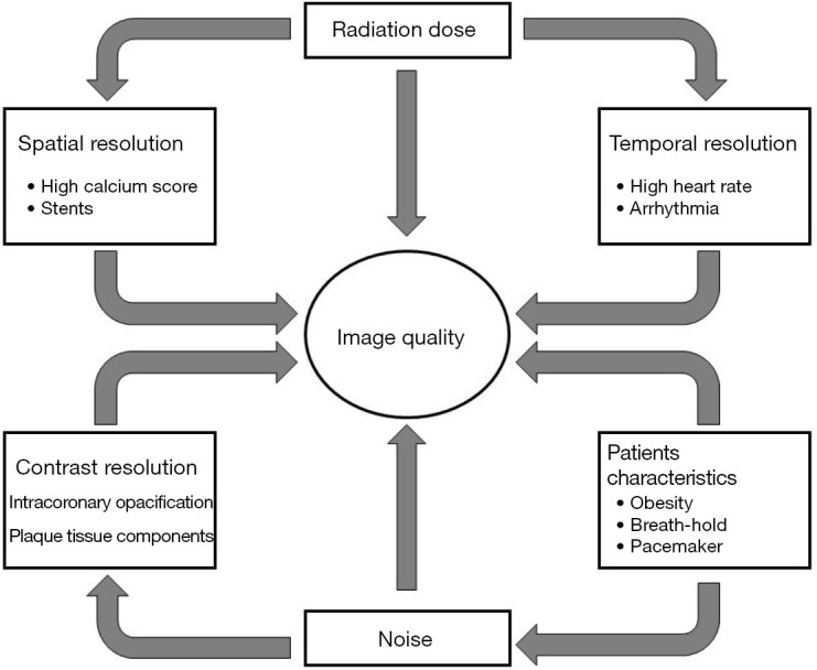
Non-invasive depiction of coronary arteries has been a great challenge for imaging specialists since the introduction of computed tomography (CT). Technological development together with improvements in spatial, temporal, and contrast resolution, progressively allowed implementation of the current clinical role of the CT assessment of coronary arteries. Several technological evolutions including hardware and software solutions of CT scanners have been developed to improve spatial and temporal resolution. The main challenges of cardiac computed tomography (CCT) are currently plaque characterization, functional assessment of stenosis and radiation dose reduction. In this review, we will discuss current standards and future improvements in CCT.

Keywords: Coronary artery disease (CAD); atherosclerosis; cardiac computed tomography (CCT); diagnosis; prognosis; therapy.


Conflict of interest statement

Conflicts of Interest: All authors have completed the ICMJE uniform disclosure form (available at http://dx.doi.org/10.21037/cdt-20-527). The series “Impact of Cardiac CT in Clinical Practice” was commissioned by the editorial office without any funding or sponsorship. FC served as the unpaid Guest Editor of the series and serves as an unpaid editorial board member of Cardiovascular Diagnosis and Therapy from Jul 2019 to Jun 2021. The authors have no other conflicts of interest to declare.

Figures

Figure 1
Figure 1
Different factors influencing and converging towards imaging quality in cardiac CT.
Figure 2
Figure 2
Case of false positive displaying intrastent restenosis at cardiac computed tomography (CCT). (A) Volume rendering; (B) multiplanar reconstruction of the left anterior descending coronary artery; (C) corresponding conventional angiogram depicting intrastent patency.
Figure 3
Figure 3
Multiplanar reconstructions of the right coronary artery displaying calcified plaques with smooth (A) and sharp filtering (B).
Figure 4
Figure 4
Cardiac CT with iterative reconstructions. (A) Volume rendering; (B) coronary artery tree map; (C,D) multiplanar reconstructions of the coronary arteries.
Figure 5
Figure 5
CT of a patient with normal coronary arteries who underwent cardiac computed tomography (CCT) with retrospective ECG-gated helical scan with ECG-controlled tube current modulation: multiplanar reconstructions of coronary arteries (A,B,C); indexed left ventricular function (D) and volume rendering (E).
Figure 6
Figure 6
Role of computed tomography in evaluation of patients potentially candidates for transcatheter aortic valve implantation (TAVI): multiplanar reconstruction of ascending aorta with its anatomic landmarks (A-E): virtual basal ring (line 1 in panel A and panel B), valve plane (line 2 in panel A and panel C), sino-tubular junction (line 3 in panel A and panel D), ascending aorta (line 4 in panel A and panel E). Panel F represents the height of left coronary ostium, panel G represents the height of right coronary ostium.
Figure 7
Figure 7
Example of FFRCT in a normal left anterior descending artery (LAD). A 55-year-old man with hypertension and atypical chest pain. (A) Normal LAD; (B) FFRCT three-dimensional model with different coronary segments FFRCT values (Software FFRct prototype by SIEMENS Frontier).
Figure 8
Figure 8
FFRCT in a diseased left anterior descending artery (LAD). A 73-year-old female with family history of cardiac heart disease, diabetes (insuline), dislipidemia, hypertension (under treatment) and typical effort angina. (A) A coronary calcified plaque in LAD, (B) an FFRCT value of 0.857872 (SmartFFR, SMARTool platform for clinical decision support system-CDSS).
Figure 9
Figure 9
Cardiac computed tomography (CCT) 3D reconstruction of the left coronary artery with plaque components: white arrows indicate calcified elements of the plaque, instead yellow arrows indicate non-calcified ones (A) and fractional flow reserve (FFR) values (B) (SmartFFR, SMARTool platform for CDSS) showing a left anterior descending coronary flow-limiting stenosis with a value of 0.599694.
Figure 10
Figure 10
Example of CT perfusion in a 36-year-old man with proximal left anterior descending artery (LAD) non calcified plaque determining segmental (sub)occlusion as indicated by the arrowhead (A) and subendocardial perfusion defect, as indicated by the arrowhead (B) during rest CCT acquisition.
Figure 11
Figure 11
Epicardial adipose tissue surrounded by the pericardium (arrows) in cardiac computed tomography (CCT): in Calcium Score (A) and angiographic phase (B).
Figure 12
Figure 12
Semi-automatic software for epicardial fat evaluation: epicardial fat delineation (A) and volume evaluation (B) in cardiac computed tomography (CCT) datasets.

References

    1. Cademartiri F, Runza G, Belgrano M, et al. Introduction to coronary imaging with 64-slice computed tomography. Radiol Med 2005;110:16-41. - PubMed
    1. Fattori R. La TC multidetettore nella diagnostica cardiovascolare. Milano, Italia: Springer, 2006.
    1. Machida H, Tanaka I, Fukui R, et al. Current and Novel Imaging Techniques in Coronary CT. RadioGraphics 2015;35:991-1010. 10.1148/rg.2015140181 - DOI - PubMed
    1. Goldman LW. Principles of CT: Multislice CT. J Nucl Med Technol 2008;36:57-68. 10.2967/jnmt.107.044826 - DOI - PubMed
    1. Wang J, Fleischmann D. Improving Spatial Resolution at CT: Development, Benefits, and Pitfalls. Radiology 2018;289:261-2. 10.1148/radiol.2018181156 - DOI - PubMed