

Extracellular Vesicles in Trypanosomatids: Host Cell Communication
- PMID: 33381465
- PMCID: PMC7767885
- DOI: 10.3389/fcimb.2020.602502
Extracellular Vesicles in Trypanosomatids: Host Cell Communication
Abstract
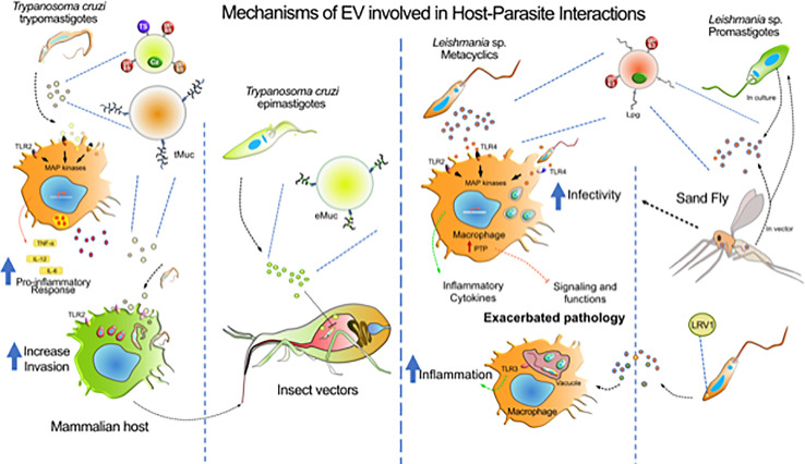
Trypanosoma cruzi, Trypanosoma brucei and Leishmania (Trypanosomatidae: Kinetoplastida) are parasitic protozoan causing Chagas disease, African Trypanosomiasis and Leishmaniases worldwide. They are vector borne diseases transmitted by triatomine bugs, Tsetse fly, and sand flies, respectively. Those diseases cause enormous economic losses and morbidity affecting not only rural and poverty areas but are also spreading to urban areas. During the parasite-host interaction, those organisms release extracellular vesicles (EVs) that are crucial for the immunomodulatory events triggered by the parasites. EVs are involved in cell-cell communication and can act as important pro-inflammatory mediators. Therefore, interface between EVs and host immune responses are crucial for the immunopathological events that those diseases exhibit. Additionally, EVs from these organisms have a role in the invertebrate hosts digestive tracts prior to parasite transmission. This review summarizes the available data on how EVs from those medically important trypanosomatids affect their interaction with vertebrate and invertebrate hosts.
Keywords: Leishmania; Trypanosoma brucei; Trypanosoma cruzi; extracellular vesicles; inflammation; innate immunity; insect vector; skin pathology.
Copyright © 2020 Torrecilhas, Soares, Schenkman, Fernández-Prada and Olivier.
Conflict of interest statement
The authors declare that the research was conducted in the absence of any commercial or financial relationships that could be construed as a potential conflict of interest.
Figures
Similar articles
-
Escaping Deleterious Immune Response in Their Hosts: Lessons from Trypanosomatids.Front Immunol. 2016 May 31;7:212. doi: 10.3389/fimmu.2016.00212. eCollection 2016. Front Immunol. 2016. PMID: 27303406 Free PMC article. Review.
-
Extracellular Vesicles during TriTryps infection: Complexity and future challenges.Mol Immunol. 2021 Apr;132:172-183. doi: 10.1016/j.molimm.2021.01.008. Epub 2021 Feb 15. Mol Immunol. 2021. PMID: 33601226 Review.
-
Comparative Analysis of Virulence Mechanisms of Trypanosomatids Pathogenic to Humans.Front Cell Infect Microbiol. 2021 Apr 16;11:669079. doi: 10.3389/fcimb.2021.669079. eCollection 2021. Front Cell Infect Microbiol. 2021. PMID: 33937106 Free PMC article. Review.
-
Impact of the Extracellular Vesicles Derived From Trypanosoma cruzi: A Paradox in Host Response and Lipid Metabolism Modulation.Front Cell Infect Microbiol. 2021 Oct 28;11:768124. doi: 10.3389/fcimb.2021.768124. eCollection 2021. Front Cell Infect Microbiol. 2021. PMID: 34778110 Free PMC article. Review.
-
Isolation and Characterization of Extracellular Vesicles Derived from Trypanosoma cruzi.Methods Mol Biol. 2019;1955:89-104. doi: 10.1007/978-1-4939-9148-8_7. Methods Mol Biol. 2019. PMID: 30868521
Cited by
-
Leishmania Vesicle-Depleted Exoproteome: What, Why, and How?Microorganisms. 2022 Dec 8;10(12):2435. doi: 10.3390/microorganisms10122435. Microorganisms. 2022. PMID: 36557688 Free PMC article. Review.
-
Host-Derived Extracellular Vesicles in Blood and Tissue Human Protozoan Infections.Microorganisms. 2023 Sep 14;11(9):2318. doi: 10.3390/microorganisms11092318. Microorganisms. 2023. PMID: 37764162 Free PMC article. Review.
-
Identification and characterization of extracellular vesicles from red cells infected with Babesia divergens and Babesia microti.Front Cell Infect Microbiol. 2022 Oct 7;12:962944. doi: 10.3389/fcimb.2022.962944. eCollection 2022. Front Cell Infect Microbiol. 2022. PMID: 36275032 Free PMC article.
-
Shedding of Trypanosoma cruzi Surface Molecules That Regulate Host Cell Invasion Involves Phospholipase C and Increases Upon Sterol Depletion.Front Cell Infect Microbiol. 2021 Oct 19;11:769722. doi: 10.3389/fcimb.2021.769722. eCollection 2021. Front Cell Infect Microbiol. 2021. PMID: 34737979 Free PMC article.
-
Structure, composition and biological properties of fungal extracellular vesicles.Microlife. 2021 Jun 24;2:uqab009. doi: 10.1093/femsml/uqab009. eCollection 2021. Microlife. 2021. PMID: 37223252 Free PMC article. Review.
References
-
- Acosta D. M., Soprano L. L., Ferrero M. R., Esteva M. I., Riarte A., Couto A. S., et al. (2012). Structural and immunological characterization of sulphatides: relevance of sulphate moieties in Trypanosoma cruzi glycoconjugates. Parasite Immunol. 34, 499–510. 10.1111/j.1365-3024.2012.01378.x - DOI - PubMed
Publication types
MeSH terms
Grants and funding
LinkOut - more resources
Full Text Sources
Medical