

Obesity Potentiates Esophageal Squamous Cell Carcinoma Growth and Invasion by AMPK-YAP Pathway
- PMID: 33381605
- PMCID: PMC7748896
- DOI: 10.1155/2020/6765474
Obesity Potentiates Esophageal Squamous Cell Carcinoma Growth and Invasion by AMPK-YAP Pathway
Abstract
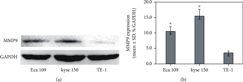
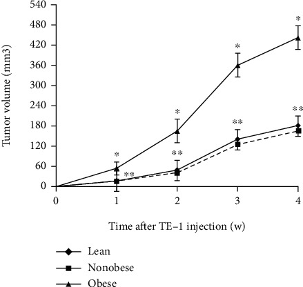
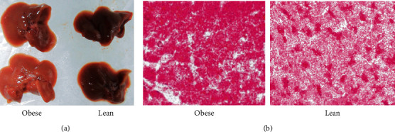
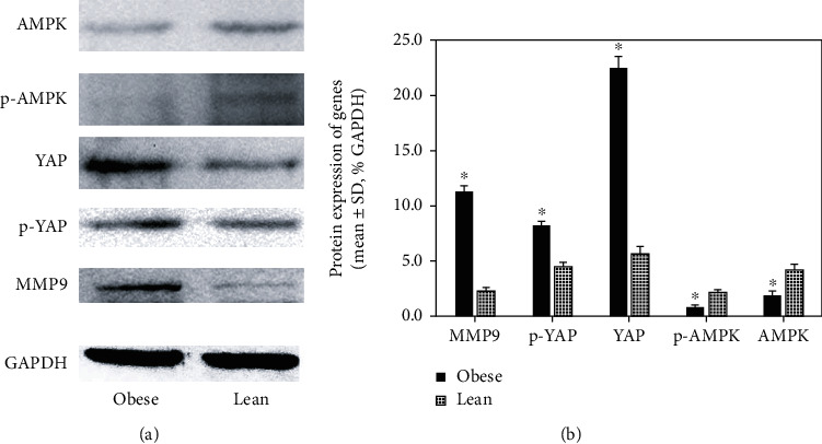
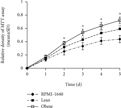
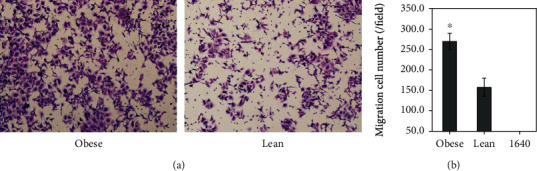
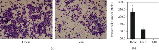
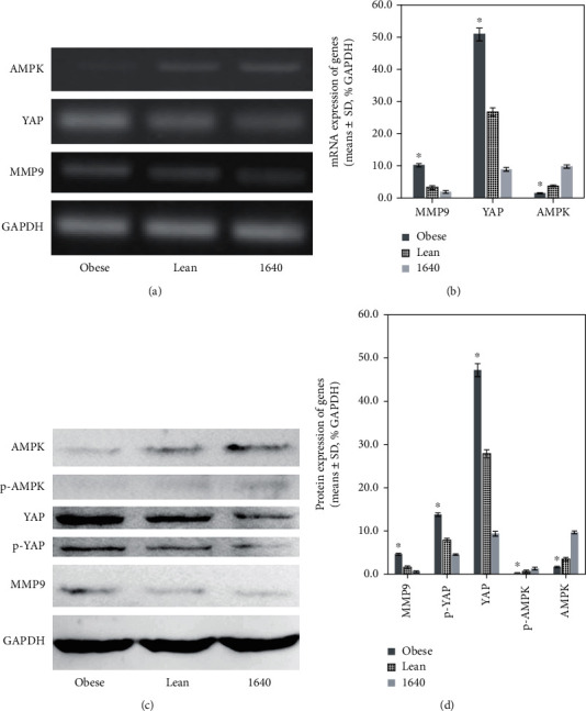
Obesity could increase the risk of esophageal squamous cell carcinoma (ESCC) and affect its growth and progression, but the mechanical links are unclear. The objective of the study was to explore the impact of obesity on ESCC growth and progression utilizing in vivo trials and cell experiments in vitro. Diet-induced obese and lean nude mice were inoculated with TE-1 cells, then studied for 4 weeks. Serum glucose, insulin, leptin, and visfatin levels were assayed. Sera of nude mice were obtained and then utilized to culture TE-1. MTT, migration and invasion assays, RT-PCR, and Western blotting were used to analyze endocrine effect of obesity on cell proliferation, migration, invasion, and related genes expression of TE-1. Obese nude mice bore larger tumor xenografts than lean animals, and were hyperglycemic and hyperinsulinemic with an elevated level of leptin and visfatin in sera, and also were accompanied by a fatty liver. As for the subcutaneous tumor xenograft model, tumors were more aggressive in obese nude mice than lean animals. Tumor weight correlated positively with mouse body weight, liver weight of mice, serum glucose, HOMA-IR, leptin, and visfatin. Obesity prompted significant TE-1 cell proliferation, migration, and invasion by endocrine mechanisms and impacted target genes. The expression of AMPK and p-AMPK protein decreased significantly (P < 0.05); MMP9, total YAP, p-YAP, and nonphosphorylated YAP protein increased significantly (P < 0.05) in the cells cultured with conditioned media and xenograft tumor from the obese group; the mRNA expression of AMPK decreased significantly (P < 0.05); YAP and MMP9 mRNA expression increased significantly (P < 0.05) in the cells exposed to conditioned media from the obese group. In conclusion, the altered adipokine milieu and metabolites in the context of obesity may promote ESCC growth in vivo; affect proliferation, migration, and invasion of ESCC cells in vitro; and regulate MMP9 and AMPK-YAP signaling pathway through complex effects including the endocrine effect.
Copyright © 2020 Jia-Huang Liu et al.
Conflict of interest statement
The authors declare that they have no conflict of interests.
Figures
Similar articles
-
Obesity and cancer: Mouse models used in studies.Front Oncol. 2023 Mar 16;13:1125178. doi: 10.3389/fonc.2023.1125178. eCollection 2023. Front Oncol. 2023. PMID: 37007087 Free PMC article. Review.
-
YAP/TEAD4-induced KIF4A contributes to the progression and worse prognosis of esophageal squamous cell carcinoma.Mol Carcinog. 2021 Jul;60(7):440-454. doi: 10.1002/mc.23303. Epub 2021 May 18. Mol Carcinog. 2021. PMID: 34003522
-
Regulation of Hippo/YAP signaling and Esophageal Squamous Carcinoma progression by an E3 ubiquitin ligase PARK2.Theranostics. 2020 Jul 25;10(21):9443-9457. doi: 10.7150/thno.46078. eCollection 2020. Theranostics. 2020. PMID: 32863938 Free PMC article.
-
Autocrine Leukemia Inhibitory Factor Promotes Esophageal Squamous Cell Carcinoma Progression via Src Family Kinase-Dependent Yes-Associated Protein Activation.Mol Cancer Res. 2020 Dec;18(12):1876-1888. doi: 10.1158/1541-7786.MCR-20-0186. Epub 2020 Oct 1. Mol Cancer Res. 2020. PMID: 33004621
-
PLCD1 Suppressed Cellular Proliferation, Invasion, and Migration via Inhibition of Wnt/β-Catenin Signaling Pathway in Esophageal Squamous Cell Carcinoma.Dig Dis Sci. 2021 Feb;66(2):442-451. doi: 10.1007/s10620-020-06218-1. Epub 2020 Mar 31. Dig Dis Sci. 2021. PMID: 32236884
Cited by
-
Multi-Region Genomic Landscape Analysis for the Preoperative Prediction of Lymph Node Metastasis in Esophageal Carcinoma.Front Genet. 2022 Mar 23;13:830601. doi: 10.3389/fgene.2022.830601. eCollection 2022. Front Genet. 2022. PMID: 35401692 Free PMC article.
-
The Good, the Bad and the New about Uric Acid in Cancer.Cancers (Basel). 2022 Oct 10;14(19):4959. doi: 10.3390/cancers14194959. Cancers (Basel). 2022. PMID: 36230882 Free PMC article. Review.
-
Dietary fat and lipid metabolism in the tumor microenvironment.Biochim Biophys Acta Rev Cancer. 2023 Nov;1878(6):188984. doi: 10.1016/j.bbcan.2023.188984. Epub 2023 Sep 16. Biochim Biophys Acta Rev Cancer. 2023. PMID: 37722512 Free PMC article. Review.
-
YAP as a therapeutic target in esophageal squamous cell carcinoma: insights and strategies.Ann Med. 2025 Dec;57(1):2536200. doi: 10.1080/07853890.2025.2536200. Epub 2025 Jul 22. Ann Med. 2025. PMID: 40693409 Free PMC article. Review.
-
Obesity and cancer: Mouse models used in studies.Front Oncol. 2023 Mar 16;13:1125178. doi: 10.3389/fonc.2023.1125178. eCollection 2023. Front Oncol. 2023. PMID: 37007087 Free PMC article. Review.
References
-
- Tian J., Zuo C., Liu G., et al. Cumulative evidence for the relationship between body mass index and the risk of esophageal cancer: an updated meta-analysis with evidence from 25 observational studies. Journal of Gastroenterology and Hepatology. 2020;35(5):730–743. doi: 10.1111/jgh.14917. - DOI - PubMed
MeSH terms
Substances
LinkOut - more resources
Full Text Sources
Medical
Research Materials
Miscellaneous
