. 2021 Jan;78(1):3-13.
doi: 10.1002/cm.21650. Epub 2021 Jan 11.

Switch-1 instability at the active site decouples ATP hydrolysis from force generation in myosin II

Affiliations

Switch-1 instability at the active site decouples ATP hydrolysis from force generation in myosin II

Benjamin C Walker et al. Cytoskeleton (Hoboken). 2021 Jan.

Abstract

Myosin active site elements (i.e., switch-1) bind both ATP and a divalent metal to coordinate ATP hydrolysis. ATP hydrolysis at the active site is linked via allosteric communication to the actin polymer binding site and lever arm movement, thus coupling the free energy of ATP hydrolysis to force generation. How active site motifs are functionally linked to actin binding and the power stroke is still poorly understood. We hypothesize that destabilizing switch-1 movement at the active site will negatively affect the tight coupling of the ATPase catalytic cycle to force production. Using a metal-switch system, we tested the effect of interfering with switch-1 coordination of the divalent metal cofactor on force generation. We found that while ATPase activity increased, motility was inhibited. Our results demonstrate that a single atom change that affects the switch-1 interaction with the divalent metal directly affects actin binding and productive force generation. Even slight modification of the switch-1 divalent metal coordination can decouple ATP hydrolysis from motility. Switch-1 movement is therefore critical for both structural communication with the actin binding site, as well as coupling the energy of ATP hydrolysis to force generation.

Keywords: RRID:SCR_002285; RRID:SCR_006643; hydrolase; kinetics; molecular motor.


Figures

FIGURE 1
FIGURE 1
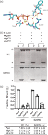
WT and S237C myosin cosedimentation with F‐actin. (a) Divalent metal coordination of the ATP hydrolysis competent state in myosin. AMPPNP from a hydrolysis competent myosin II structure (PDB: 3MYK) was replaced with ATP from another myosin structure (PDB: 1FMW), after alignment using the P‐loop residues. The magnesium ion is shown as a green sphere and water molecules are shown as red spheres. The yellow dotted lines represent polar contacts. (b) Representative Coomassie Blue‐stained SDS‐PAGE gel of WT or S237C myosin (1 μM) binding to phalloidin‐stabilized F‐actin (1.5 μM). Equivalent volumes of the supernatant (S) and pellet (P) fractions were electrophoresed from each reaction and quantified by densitometry (see panel C and table). Myosin is indicated with m and actin with a. (c) Bar graph and table representing the mean fraction ± SD of WT (n = 5) and S237C (n = 3) myosin bound (f b) to F‐actin [Color figure can be viewed at wileyonlinelibrary.com]
FIGURE 2
FIGURE 2
Steady‐state ATPase activity of WT and S237C myosin. The NADH‐coupled assay was used to determine the ATPase activity of 0.3 μM WT or S237C myosin with 1 mM ATP and 1 mM divalent metal (Mg2+, Mn2+, or Ca2+) in the presence or absence of 20 μM phalloidin‐stabilized F‐actin (n = 3). (a) Bar graph of the mean ATP turnover rates ± SD with different divalent metals. Unpaired t‐tests were performed for each condition ± actin. **, p ≤ .01; ***, p ≤ .001. (b) Table of the mean ± SD rate of ATP turnover for each myosin and condition
FIGURE 3
FIGURE 3
F‐actin gliding by WT and S237C myosin under various metal conditions. (a) Schematic showing myosin immobilized on a microscope glass chamber and FITC‐labeled F‐actin bound to myosin before addition of divalent metal and ATP to induce gliding. (b) Representative kymographs of WT and S237C bound F‐actin filaments in the different ATP and divalent metal compositions. (c) Scatterplot of the gliding velocity of individual actin filaments with the mean ± SD indicated. For each condition, gliding velocities were calculated from the slope of 10 filament kymographs per gliding chamber from five separate experiments (n = 50). WT(MgATP) = 0.11 ± 0.01 μm s−1, WT(MnATP) = 0.04 ± 0.01 μm s−1. For all other conditions gliding was not detected. An unpaired t‐test was performed to test significance between the indicated conditions. ****p ≤ .0001. (d) F‐actin gliding velocity of individual actin filaments with the mean ± SD indicated of WT with and without S237C under MgATP (n = 2) and MnATP (n = 3) conditions. A minimum of 10 F‐actin filaments were tracked per condition per experiment. Experiments were performed as described with WT and S237C mixtures varying from 1:1 to 5:2 ratios. WT(MgATP) = 0.11 ± 0.02 μm s−1, WT(MnATP) = 0.04 ± 0.01 μm s−1. For all other conditions gliding was not detected. An unpaired t‐test was performed to test significance between the indicated conditions. ****p ≤ .0001. (e) F‐actin gliding chambers were filled with the NADH‐coupled system and incubated at 25°C for 45 min before the solution was extracted and the amount of ADP produced measured. The amount of ADP produced in each chamber is graphed as a scatter plot with the mean ± SD indicated. WT(MgATP) = 2 ± 6 μM (n = 4), WT(MnATP) = 20 ± 13 μM (n = 5), S237C(MgATP) = 181 ± 88 μM (n = 4), S237C(MnATP) = 34 ± 16 μM (n = 5). An unpaired t‐test was performed to test significance between the indicated conditions. *p < .05 [Color figure can be viewed at wileyonlinelibrary.com]
FIGURE 4
FIGURE 4
Models of ATPase cycle and force generation for myosin II. Kinetic models for myosin II motor constructs (gray) as they bind and hydrolyze ATP and release products in the presence of F‐actin filaments (green). Main kinetic pathways are highlighted. The major portion of the power stroke occurs with phosphate release and is highlighted by a curved arrow. (a) Model for the WT MgATPase cycle. The conformational change that leads to tight F‐actin binding intermediate, the power stroke, and P i release limits both the basal and F‐actin stimulated cycles (Gyimesi et al., 2008; Stein, Chock, & Eisenberg, 1984). Therefore, the dominant species in the ATPase cycle is the weak ADP‐P i state, and the power stroke occurs primarily when bound to F‐actin (productive). (b) Model for the WT and S237C MnATPase cycles. The conformational changes that are associated with P i and ADP release limit the basal cycle while only the conformational change associated with P i release limits the actomyosin cycle. Consequently, a significant fraction will undergo the power stroke off the filament (futile). We suggest that ADP release is hyper strain sensitive in the S237C and leads to inhibition of force generation along the F‐actin filament [Color figure can be viewed at wileyonlinelibrary.com]

References

    1. Anson, M. , Geeves, M. A. , Kurzawa, S. E. , & Manstein, D. J. (1996). Myosin motors with artificial lever arms. The EMBO Journal, 15(22), 6069–6074. 10.1002/j.1460-2075.1996.tb00995.x - DOI - PMC - PubMed
    1. Bloemink, M. J. , & Geeves, M. A. (2011). Shaking the myosin family tree: Biochemical kinetics defines four types of myosin motor. Seminars in Cell & Developmental Biology, 22(9), 961–967. 10.1016/j.semcdb.2011.09.015 - DOI - PMC - PubMed
    1. Bobkov, A. A. , Sutoh, K. , & Reisler, E. (1997). Nucleotide and actin binding properties of the isolated motor domain from Dictyostelium discoideum myosin. Journal of Muscle Research and Cell Motility, 18(5), 563–571. 10.1023/a:1018667319386 - DOI - PubMed
    1. Brizendine, R. K. , Sheehy, G. G. , Alcala, D. B. , Novenschi, S. I. , Baker, J. E. , & Cremo, C. R. (2017). A mixed‐kinetic model describes unloaded velocities of smooth, skeletal, and cardiac muscle myosin filaments in vitro. Science Advances, 3(12), eaao2267. 10.1126/sciadv.aao2267 - DOI - PMC - PubMed
    1. Burke, M. , Reisler, E. , & Harrington, W. F. (1973). Myosin ATP hydrolysis: A mechanism involving a magnesium chelate complex. Proceedings of the National Academy of Sciences of the United States of America, 70(12), 3793–3796. 10.1073/pnas.70.12.3793 - DOI - PMC - PubMed

Publication types

LinkOut - more resources